 Movie
Movie Controller


+ Open data

- Basic information
| Entry | Database: PDB / ID: 1iap | ||||||
|---|---|---|---|---|---|---|---|
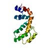
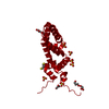
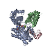
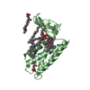
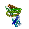
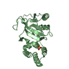
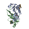
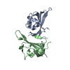
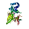
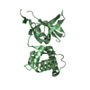
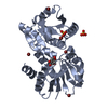
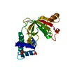
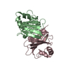
| Title | CRYSTAL STRUCTURE OF P115RHOGEF RGRGS DOMAIN | ||||||
 Components Components | GUANINE NUCLEOTIDE EXCHANGE FACTOR P115RHOGEF | ||||||
 Keywords Keywords | SIGNALING PROTEIN / p115 / RhoGEF / RGS / RGRGS | ||||||
| Function / homology |  Function and homology information Function and homology informationRho-activating G protein-coupled receptor signaling pathway / regulation of small GTPase mediated signal transduction / RHOB GTPase cycle / NRAGE signals death through JNK / RHOC GTPase cycle / RHOA GTPase cycle / Rho protein signal transduction / guanyl-nucleotide exchange factor activity / GTPase activator activity / G protein-coupled receptor binding ...Rho-activating G protein-coupled receptor signaling pathway / regulation of small GTPase mediated signal transduction / RHOB GTPase cycle / NRAGE signals death through JNK / RHOC GTPase cycle / RHOA GTPase cycle / Rho protein signal transduction / guanyl-nucleotide exchange factor activity / GTPase activator activity / G protein-coupled receptor binding / G alpha (12/13) signalling events / G protein-coupled receptor signaling pathway / RNA binding / plasma membrane / cytoplasm / cytosol Similarity search - Function | ||||||
| Biological species |  Homo sapiens (human) Homo sapiens (human) | ||||||
| Method |  X-RAY DIFFRACTION / SYNCHROTRON / MOLECULAR REPLACEMENT / Resolution: 1.9 Å X-RAY DIFFRACTION / SYNCHROTRON / MOLECULAR REPLACEMENT / Resolution: 1.9 Å | ||||||
 Authors Authors | Sprang, S.R. / Chen, Z. | ||||||
 Citation Citation | Journal: Nat.Struct.Biol. / Year: 2001 Title: Structure of the rgRGS domain of p115RhoGEF. Authors: Chen, Z. / Wells, C.D. / Sternweis, P.C. / Sprang, S.R. | ||||||
| History |
|
- Structure visualization
| Structure viewer | Molecule: MolmilJmol/JSmol |
|---|
- Downloads & links
-Download
| PDBx/mmCIF format | 1iap.cif.gz | 51.9 KB | Display | PDBx/mmCIF format |
|---|---|---|---|---|
| PDB format | pdb1iap.ent.gz | 37.1 KB | Display | PDB format |
| PDBx/mmJSON format | 1iap.json.gz | Tree view | PDBx/mmJSON format | |
| Others |  Other downloads Other downloads |
-Validation report
| Arichive directory | https://data.pdbj.org/pub/pdb/validation_reports/ia/1iapftp://data.pdbj.org/pub/pdb/validation_reports/ia/1iap | HTTPS FTP |
|---|
-Related structure data
| Related structure data | |
|---|---|
| Similar structure data |
-Links
 PDBj
PDBj


- Assembly
| Deposited unit | 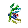
| ||||||||
|---|---|---|---|---|---|---|---|---|---|
| 1 |
| ||||||||
| Unit cell |
|
-Components
| #1: Protein | Mass: 24276.924 Da / Num. of mol.: 1 / Fragment: N-TERMINAL RGS DOMAIN Source method: isolated from a genetically manipulated source Source: (gene. exp.) Homo sapiens (human) / Plasmid: PTRCC / Species (production host): Escherichia coli / Production host:  |
|---|---|
| #2: Water | ChemComp-HOH /  Mass: 18.015 Da / Num. of mol.: 91 / Source method: isolated from a natural source / Formula: H2O Mass: 18.015 Da / Num. of mol.: 91 / Source method: isolated from a natural source / Formula: H2O |
-Experimental details
-Experiment
| Experiment | Method: X-RAY DIFFRACTION / Number of used crystals: 1 |
|---|
- Sample preparation
| Crystal | Density Matthews: 1.73 Å3/Da / Density % sol: 28.3 % | ||||||||||||||||||||
|---|---|---|---|---|---|---|---|---|---|---|---|---|---|---|---|---|---|---|---|---|---|
| Crystal grow | Temperature: 293 K / Method: vapor diffusion, hanging drop / pH: 9.5 Details: PEG 3350, ammonium formate, CHES, pH 9.5, VAPOR DIFFUSION, HANGING DROP, temperature 293.0K | ||||||||||||||||||||
| Crystal grow | *PLUS Temperature: 20 ℃ / Method: vapor diffusion / PH range low: 9.6 / PH range high: 9.3 | ||||||||||||||||||||
| Components of the solutions | *PLUS
|
-Data collection
| Diffraction | Mean temperature: 100 K |
|---|---|
| Diffraction source | Source: SYNCHROTRON / Site: ALS  / Beamline: 5.0.2 / Wavelength: 0.976 Å / Beamline: 5.0.2 / Wavelength: 0.976 Å |
| Detector | Type: ADSC QUANTUM 4 / Detector: CCD / Date: Jan 1, 2001 |
| Radiation | Protocol: SINGLE WAVELENGTH / Monochromatic (M) / Laue (L): M / Scattering type: x-ray |
| Radiation wavelength | Wavelength: 0.976 Å / Relative weight: 1 |
| Reflection | Resolution: 1.9→16 Å / Num. all: 14781 / Num. obs: 13303 / % possible obs: 90 % / Observed criterion σ(F): 0 / Observed criterion σ(I): 0 / Redundancy: 3.8 % / Biso Wilson estimate: 18.6 Å2 / Rmerge(I) obs: 0.12 / Rsym value: 0.12 / Net I/σ(I): 12.1 |
| Reflection shell | Resolution: 1.9→1.97 Å / Redundancy: 3.4 % / Rmerge(I) obs: 0.485 / Mean I/σ(I) obs: 4.8 / Num. unique all: 1152 / Rsym value: 0.485 / % possible all: 79.6 |
| Reflection shell | *PLUS % possible obs: 79.6 % |
- Processing
| Software |
| |||||||||||||||||||||||||
|---|---|---|---|---|---|---|---|---|---|---|---|---|---|---|---|---|---|---|---|---|---|---|---|---|---|---|
| Refinement | Method to determine structure: MOLECULAR REPLACEMENT Starting model: SE-MET STRUCTURE OF THE SAME PROTEIN SOLVED BY MAD PHASING FROM A FOUR-WAVELENGTH MAD DATASET COLLECTED AT CHESS F2 BEAMLINE (UNPUBLISHED DATA) Resolution: 1.9→15.55 Å / Rfactor Rfree error: 0.007 / Data cutoff high absF: 1423020.44 / Data cutoff low absF: 0 / Isotropic thermal model: RESTRAINED / Cross valid method: THROUGHOUT / σ(F): 0 / σ(I): 0 / Stereochemistry target values: Engh & Huber
| |||||||||||||||||||||||||
| Solvent computation | Solvent model: FLAT MODEL / Bsol: 48.57 Å2 / ksol: 0.364 e/Å3 | |||||||||||||||||||||||||
| Displacement parameters | Biso mean: 33 Å2
| |||||||||||||||||||||||||
| Refine analyze |
| |||||||||||||||||||||||||
| Refinement step | Cycle: LAST / Resolution: 1.9→15.55 Å
| |||||||||||||||||||||||||
| Refine LS restraints |
| |||||||||||||||||||||||||
| LS refinement shell | Resolution: 1.9→2.02 Å / Rfactor Rfree error: 0.025 / Total num. of bins used: 6
| |||||||||||||||||||||||||
| Xplor file |
| |||||||||||||||||||||||||
| Software | *PLUS Name: CNS / Version: 0.9 / Classification: refinement | |||||||||||||||||||||||||
| Refinement | *PLUS σ(F): 0 / % reflection Rfree: 10.3 % | |||||||||||||||||||||||||
| Solvent computation | *PLUS | |||||||||||||||||||||||||
| Displacement parameters | *PLUS Biso mean: 33 Å2 | |||||||||||||||||||||||||
| Refine LS restraints | *PLUS
| |||||||||||||||||||||||||
| LS refinement shell | *PLUS Rfactor Rfree: 0.339 / % reflection Rfree: 9.4 % / Rfactor Rwork: 0.304 |
