 Movie
Movie Controller

[English] 日本語
 Yorodumi
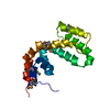
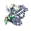
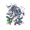
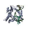
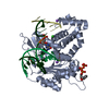
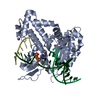
- PDB-1fqj: CRYSTAL STRUCTURE OF THE HETEROTRIMERIC COMPLEX OF THE RGS DOMAIN... -
+ Open data

- Basic information
| Entry | Database: PDB / ID: 1fqj | ||||||
|---|---|---|---|---|---|---|---|
| Title | CRYSTAL STRUCTURE OF THE HETEROTRIMERIC COMPLEX OF THE RGS DOMAIN OF RGS9, THE GAMMA SUBUNIT OF PHOSPHODIESTERASE AND THE GT/I1 CHIMERA ALPHA SUBUNIT [(RGS9)-(PDEGAMMA)-(GT/I1ALPHA)-(GDP)-(ALF4-)-(MG2+)] | ||||||
 Components Components |
| ||||||
 Keywords Keywords | SIGNALING PROTEIN / RGS9 / transducin / effector / PDEgamma / G Protein / Phototransduction / rod / RGS / phosphodiesterase / GAP | ||||||
| Function / homology |  Function and homology information Function and homology informationnegative regulation of cyclic-nucleotide phosphodiesterase activity / adenylate cyclase regulator activity / Extra-nuclear estrogen signaling / Adenylate cyclase inhibitory pathway / 3',5'-cyclic-GMP phosphodiesterase / detection of light stimulus involved in visual perception / G protein-coupled receptor complex / Inactivation, recovery and regulation of the phototransduction cascade / Activation of the phototransduction cascade / positive regulation of G protein-coupled receptor signaling pathway ...negative regulation of cyclic-nucleotide phosphodiesterase activity / adenylate cyclase regulator activity / Extra-nuclear estrogen signaling / Adenylate cyclase inhibitory pathway / 3',5'-cyclic-GMP phosphodiesterase / detection of light stimulus involved in visual perception / G protein-coupled receptor complex / Inactivation, recovery and regulation of the phototransduction cascade / Activation of the phototransduction cascade / positive regulation of G protein-coupled receptor signaling pathway / regulation of G protein-coupled receptor signaling pathway / negative regulation of synaptic transmission / Adrenaline,noradrenaline inhibits insulin secretion / ADP signalling through P2Y purinoceptor 12 / GTPase activating protein binding / G alpha (i) signalling events / G protein-coupled dopamine receptor signaling pathway / Ca2+ pathway / photoreceptor outer segment membrane / G alpha (i) signalling events / neurotransmitter receptor localization to postsynaptic specialization membrane / cGMP binding / 3',5'-cyclic-GMP phosphodiesterase activity / acyl binding / phototransduction, visible light / phototransduction / response to light stimulus / negative regulation of signal transduction / positive regulation of epidermal growth factor receptor signaling pathway / adenylate cyclase inhibitor activity / positive regulation of protein localization to cell cortex / photoreceptor inner segment / T cell migration / visual perception / D2 dopamine receptor binding / adenylate cyclase-inhibiting serotonin receptor signaling pathway / G protein-coupled serotonin receptor binding / cellular response to forskolin / regulation of mitotic spindle organization / GTPase activator activity / chemokine-mediated signaling pathway / response to prostaglandin E / positive regulation of cholesterol biosynthetic process / negative regulation of insulin secretion / G protein-coupled receptor binding / centriolar satellite / G-protein beta/gamma-subunit complex binding / adenylate cyclase-modulating G protein-coupled receptor signaling pathway / adenylate cyclase-inhibiting G protein-coupled receptor signaling pathway / photoreceptor disc membrane / GDP binding / heterotrimeric G-protein complex / G protein activity / midbody / cell cortex / molecular adaptor activity / Hydrolases; Acting on acid anhydrides; Acting on GTP to facilitate cellular and subcellular movement / positive regulation of MAPK cascade / neuron projection / intracellular signal transduction / postsynapse / ciliary basal body / G protein-coupled receptor signaling pathway / cell division / GTPase activity / centrosome / protein kinase binding / GTP binding / nucleolus / glutamatergic synapse / magnesium ion binding / Golgi apparatus / protein-containing complex / zinc ion binding / nucleoplasm / metal ion binding / plasma membrane / cytoplasm / cytosol Similarity search - Function | ||||||
| Biological species |  | ||||||
| Method |  X-RAY DIFFRACTION / SYNCHROTRON / Resolution: 2.02 Å X-RAY DIFFRACTION / SYNCHROTRON / Resolution: 2.02 Å | ||||||
 Authors Authors | Slep, K.C. / Kercher, M.A. / He, W. / Cowan, C.W. / Wensel, T.G. / Sigler, P.B. | ||||||
 Citation Citation | Journal: Nature / Year: 2001 Title: Structural determinants for regulation of phosphodiesterase by a G protein at 2.0 A. Authors: Slep, K.C. / Kercher, M.A. / He, W. / Cowan, C.W. / Wensel, T.G. / Sigler, P.B. | ||||||
| History |
|
- Structure visualization
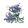
| Structure viewer | Molecule: MolmilJmol/JSmol |
|---|
- Downloads & links
-Download
| PDBx/mmCIF format | 1fqj.cif.gz | 213 KB | Display | PDBx/mmCIF format |
|---|---|---|---|---|
| PDB format | pdb1fqj.ent.gz | 169 KB | Display | PDB format |
| PDBx/mmJSON format | 1fqj.json.gz | Tree view | PDBx/mmJSON format | |
| Others |  Other downloads Other downloads |
-Validation report
| Arichive directory | https://data.pdbj.org/pub/pdb/validation_reports/fq/1fqjftp://data.pdbj.org/pub/pdb/validation_reports/fq/1fqj | HTTPS FTP |
|---|
-Related structure data


-Links
 PDBj
PDBj


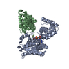
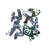
- Assembly
| Deposited unit | 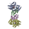
| ||||||||
|---|---|---|---|---|---|---|---|---|---|
| 1 | 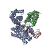
| ||||||||
| 2 | 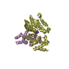
| ||||||||
| Unit cell |
|
-Components
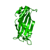
-Protein , 2 types, 4 molecules ADBE
| #1: Protein | Mass: 37361.656 Da / Num. of mol.: 2 Fragment: UNP P04695 residues 26-215 and 295-350 linked via UNP P10824 residues 220-298 Source method: isolated from a genetically manipulated source Details: THE CHIMERA COMPRISES RESIDUES 26 TO 215 OF BOVINE GT, RESIDUES 220 TO 298 OF RAT GI1, AND RESIDUES 295 TO 350 OF BOVINE GT Source: (gene. exp.) Genus: Bos, Rattus / Species: , / Gene: GNAT1, Gnai1, Gnai-1 / Plasmid: PHIS6(T7) / Production host:  #2: Protein | Mass: 17419.176 Da / Num. of mol.: 2 / Fragment: RGS DOMAIN UNP RESIDUES 276-422 Source method: isolated from a genetically manipulated source Source: (gene. exp.) |
|---|
-Protein/peptide , 1 types, 1 molecules C
| #3: Protein/peptide | Mass: 4559.094 Da / Num. of mol.: 1 / Fragment: UNP RESIDUES 46-87 Source method: isolated from a genetically manipulated source Source: (gene. exp.) References: UniProt: P04972, 3',5'-cyclic-GMP phosphodiesterase |
|---|
-Non-polymers , 4 types, 362 molecules 


| #4: Chemical |  Mass: 24.305 Da / Num. of mol.: 2 / Source method: obtained synthetically / Formula: Mg Mass: 24.305 Da / Num. of mol.: 2 / Source method: obtained synthetically / Formula: Mg#5: Chemical |  Mass: 102.975 Da / Num. of mol.: 2 / Source method: obtained synthetically / Formula: AlF4 Mass: 102.975 Da / Num. of mol.: 2 / Source method: obtained synthetically / Formula: AlF4#6: Chemical |  Type: RNA linking / Mass: 443.201 Da / Num. of mol.: 2 / Source method: obtained synthetically / Formula: C10H15N5O11P2 / Comment: GDP, energy-carrying molecule*YM Type: RNA linking / Mass: 443.201 Da / Num. of mol.: 2 / Source method: obtained synthetically / Formula: C10H15N5O11P2 / Comment: GDP, energy-carrying molecule*YM#7: Water | ChemComp-HOH / | Mass: 18.015 Da / Num. of mol.: 356 / Source method: isolated from a natural source / Formula: H2O |
|---|
-Experimental details
-Experiment
| Experiment | Method: X-RAY DIFFRACTION / Number of used crystals: 1 |
|---|
- Sample preparation
| Crystal | Density Matthews: 3 Å3/Da / Density % sol: 59 % | ||||||||||||||||||||||||||||||||||||
|---|---|---|---|---|---|---|---|---|---|---|---|---|---|---|---|---|---|---|---|---|---|---|---|---|---|---|---|---|---|---|---|---|---|---|---|---|---|
| Crystal grow | Temperature: 277 K / Method: vapor diffusion, hanging drop / pH: 9 Details: 15.0% PEG8000, 200mM Tris pH9.0, 0.2% beta-ME, 1mM (NH4)2WS4, VAPOR DIFFUSION, HANGING DROP, temperature 277K | ||||||||||||||||||||||||||||||||||||
| Crystal grow | *PLUS Temperature: 4 ℃ / pH: 8 | ||||||||||||||||||||||||||||||||||||
| Components of the solutions | *PLUS
|
-Data collection
| Diffraction | Mean temperature: 100 K |
|---|---|
| Diffraction source | Source: SYNCHROTRON / Site: APS  / Beamline: 19-ID / Wavelength: 0.9793 / Beamline: 19-ID / Wavelength: 0.9793 |
| Detector | Type: APS-1 / Detector: CCD / Date: Sep 6, 1999 |
| Radiation | Protocol: SINGLE WAVELENGTH / Monochromatic (M) / Laue (L): M / Scattering type: x-ray |
| Radiation wavelength | Wavelength: 0.9793 Å / Relative weight: 1 |
| Reflection | Resolution: 2.02→50 Å / Num. all: 2227054 / Num. obs: 93531 / % possible obs: 99.8 % / Observed criterion σ(F): 0 / Observed criterion σ(I): 0 / Redundancy: 6.7 % / Biso Wilson estimate: 8.5 Å2 / Rmerge(I) obs: 0.09 / Net I/σ(I): 19.5 |
| Reflection shell | Resolution: 2.02→2.09 Å / Redundancy: 6.1 % / Rmerge(I) obs: 0.553 / Num. unique all: 2094 / % possible all: 99.7 |
| Reflection | *PLUS Num. measured all: 2227054 / Rmerge(I) obs: 0.09 |
| Reflection shell | *PLUS % possible obs: 99.7 % / Mean I/σ(I) obs: 4.4 |
- Processing
| Software |
| ||||||||||||||||||||||||||||||||||||||||||||||||||||||||||||
|---|---|---|---|---|---|---|---|---|---|---|---|---|---|---|---|---|---|---|---|---|---|---|---|---|---|---|---|---|---|---|---|---|---|---|---|---|---|---|---|---|---|---|---|---|---|---|---|---|---|---|---|---|---|---|---|---|---|---|---|---|---|
| Refinement | Resolution: 2.02→50 Å / Rfactor Rfree error: 0.002 / Data cutoff high absF: 2012134.44 / Data cutoff low absF: 0 / Isotropic thermal model: OVERALL / Cross valid method: THROUGHOUT / σ(F): 0 / σ(I): 0 / Stereochemistry target values: Engh & Huber
| ||||||||||||||||||||||||||||||||||||||||||||||||||||||||||||
| Solvent computation | Solvent model: FLAT MODEL / Bsol: 35.33 Å2 / ksol: 0.344 e/Å3 | ||||||||||||||||||||||||||||||||||||||||||||||||||||||||||||
| Displacement parameters | Biso mean: 33.8 Å2
| ||||||||||||||||||||||||||||||||||||||||||||||||||||||||||||
| Refine analyze |
| ||||||||||||||||||||||||||||||||||||||||||||||||||||||||||||
| Refinement step | Cycle: LAST / Resolution: 2.02→50 Å
| ||||||||||||||||||||||||||||||||||||||||||||||||||||||||||||
| Refine LS restraints |
| ||||||||||||||||||||||||||||||||||||||||||||||||||||||||||||
| LS refinement shell | Resolution: 2.02→2.15 Å / Rfactor Rfree error: 0.007 / Total num. of bins used: 6
| ||||||||||||||||||||||||||||||||||||||||||||||||||||||||||||
| Xplor file |
| ||||||||||||||||||||||||||||||||||||||||||||||||||||||||||||
| Software | *PLUS Name: CNS / Version: 1 / Classification: refinement | ||||||||||||||||||||||||||||||||||||||||||||||||||||||||||||
| Refinement | *PLUS Lowest resolution: 50 Å / σ(F): 0 / % reflection Rfree: 9.8 % | ||||||||||||||||||||||||||||||||||||||||||||||||||||||||||||
| Solvent computation | *PLUS | ||||||||||||||||||||||||||||||||||||||||||||||||||||||||||||
| Displacement parameters | *PLUS Biso mean: 33.8 Å2 | ||||||||||||||||||||||||||||||||||||||||||||||||||||||||||||
| Refine LS restraints | *PLUS
| ||||||||||||||||||||||||||||||||||||||||||||||||||||||||||||
| LS refinement shell | *PLUS Rfactor Rfree: 0.342 / % reflection Rfree: 9.9 % / Rfactor Rwork: 0.306 / Rfactor obs: 0.319 |
