 Movie
Movie Controller


+ Open data

- Basic information
| Entry | Database: PDB / ID: 1agr | ||||||
|---|---|---|---|---|---|---|---|
| Title | COMPLEX OF ALF4-ACTIVATED GI-ALPHA-1 WITH RGS4 | ||||||
 Components Components |
| ||||||
 Keywords Keywords | COMPLEX (SIGNAL TRANSDUCTION/REGULATOR) / GI-ALPHA-1 / HYDROLASE / SIGNAL TRANSDUCTION / RGS4 / COMPLEX (SIGNAL TRANSDUCTION-REGULATOR) / GTP-BINDING / GTPASE ACTIVATING PROTEIN / COMPLEX (SIGNAL TRANSDUCTION-REGULATOR) complex | ||||||
| Function / homology |  Function and homology information Function and homology informationnegative regulation of glycine import across plasma membrane / negative regulation of dopamine receptor signaling pathway / adenylate cyclase regulator activity / Extra-nuclear estrogen signaling / Adenylate cyclase inhibitory pathway / negative regulation of potassium ion transmembrane transport / dorsal root ganglion development / negative regulation of cell growth involved in cardiac muscle cell development / regulation of actin filament organization / regulation of potassium ion transmembrane transport ...negative regulation of glycine import across plasma membrane / negative regulation of dopamine receptor signaling pathway / adenylate cyclase regulator activity / Extra-nuclear estrogen signaling / Adenylate cyclase inhibitory pathway / negative regulation of potassium ion transmembrane transport / dorsal root ganglion development / negative regulation of cell growth involved in cardiac muscle cell development / regulation of actin filament organization / regulation of potassium ion transmembrane transport / negative regulation of synaptic transmission / Adrenaline,noradrenaline inhibits insulin secretion / ADP signalling through P2Y purinoceptor 12 / G alpha (q) signalling events / GTPase activating protein binding / G alpha (i) signalling events / negative regulation of G protein-coupled receptor signaling pathway / neurotransmitter receptor localization to postsynaptic specialization membrane / regulation of calcium ion transport / G-protein alpha-subunit binding / positive regulation of heart rate / adenylate cyclase inhibitor activity / positive regulation of protein localization to cell cortex / T cell migration / D2 dopamine receptor binding / adenylate cyclase-inhibiting serotonin receptor signaling pathway / G protein-coupled serotonin receptor binding / cellular response to forskolin / positive regulation of excitatory postsynaptic potential / regulation of mitotic spindle organization / GTPase activator activity / chemokine-mediated signaling pathway / response to amphetamine / response to cocaine / response to prostaglandin E / positive regulation of cholesterol biosynthetic process / negative regulation of insulin secretion / G protein-coupled receptor binding / centriolar satellite / G-protein beta/gamma-subunit complex binding / adenylate cyclase-modulating G protein-coupled receptor signaling pathway / adenylate cyclase-inhibiting G protein-coupled receptor signaling pathway / GDP binding / heterotrimeric G-protein complex / G protein activity / midbody / cell cortex / Hydrolases; Acting on acid anhydrides; Acting on GTP to facilitate cellular and subcellular movement / response to ethanol / postsynapse / ciliary basal body / G protein-coupled receptor signaling pathway / cell division / GTPase activity / centrosome / protein kinase binding / GTP binding / nucleolus / glutamatergic synapse / magnesium ion binding / Golgi apparatus / protein-containing complex / nucleoplasm / nucleus / plasma membrane / cytoplasm / cytosol Similarity search - Function | ||||||
| Biological species |  | ||||||
| Method |  X-RAY DIFFRACTION / SYNCHROTRON / MOLECULAR REPLACEMENT, DENSITY MODIFICATION / Resolution: 2.8 Å X-RAY DIFFRACTION / SYNCHROTRON / MOLECULAR REPLACEMENT, DENSITY MODIFICATION / Resolution: 2.8 Å | ||||||
 Authors Authors | Tesmer, J.J.G. / Sprang, S.R. | ||||||
 Citation Citation | Journal: Cell(Cambridge,Mass.) / Year: 1997 Title: Structure of RGS4 bound to AlF4--activated G(i alpha1): stabilization of the transition state for GTP hydrolysis. Authors: Tesmer, J.J. / Berman, D.M. / Gilman, A.G. / Sprang, S.R. | ||||||
| History |
|
- Structure visualization
| Structure viewer | Molecule: MolmilJmol/JSmol |
|---|
- Downloads & links
-Download
| PDBx/mmCIF format | 1agr.cif.gz | 206.3 KB | Display | PDBx/mmCIF format |
|---|---|---|---|---|
| PDB format | pdb1agr.ent.gz | 163 KB | Display | PDB format |
| PDBx/mmJSON format | 1agr.json.gz | Tree view | PDBx/mmJSON format | |
| Others |  Other downloads Other downloads |
-Validation report
| Arichive directory | https://data.pdbj.org/pub/pdb/validation_reports/ag/1agrftp://data.pdbj.org/pub/pdb/validation_reports/ag/1agr | HTTPS FTP |
|---|
-Related structure data
| Related structure data | 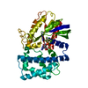 1gfiS S: Starting model for refinement |
|---|---|
| Similar structure data |


-Links
 PDBj
PDBj


- Assembly
| Deposited unit | 
| ||||||||
|---|---|---|---|---|---|---|---|---|---|
| 1 | 
| ||||||||
| 2 | 
| ||||||||
| Unit cell |
| ||||||||
| Noncrystallographic symmetry (NCS) | NCS oper: (Code: given Matrix: (0.964372, 0.208506, -0.162827), Vector: |
-Components
-Protein , 2 types, 4 molecules ADEH
| #1: Protein | Mass: 40267.836 Da / Num. of mol.: 2 / Fragment: ALPHA-1 Source method: isolated from a genetically manipulated source Source: (gene. exp.)  #2: Protein | Mass: 23286.697 Da / Num. of mol.: 2 Source method: isolated from a genetically manipulated source Source: (gene. exp.) |
|---|
-Non-polymers , 5 types, 66 molecules 


| #3: Chemical |  Mass: 24.305 Da / Num. of mol.: 2 / Source method: obtained synthetically / Formula: Mg Mass: 24.305 Da / Num. of mol.: 2 / Source method: obtained synthetically / Formula: Mg#4: Chemical |  Mass: 102.975 Da / Num. of mol.: 2 / Source method: obtained synthetically / Formula: AlF4 Mass: 102.975 Da / Num. of mol.: 2 / Source method: obtained synthetically / Formula: AlF4#5: Chemical |  Type: RNA linking / Mass: 443.201 Da / Num. of mol.: 2 / Source method: obtained synthetically / Formula: C10H15N5O11P2 / Comment: GDP, energy-carrying molecule*YM Type: RNA linking / Mass: 443.201 Da / Num. of mol.: 2 / Source method: obtained synthetically / Formula: C10H15N5O11P2 / Comment: GDP, energy-carrying molecule*YM#6: Chemical |  Mass: 192.124 Da / Num. of mol.: 2 / Source method: obtained synthetically / Formula: C6H8O7 Mass: 192.124 Da / Num. of mol.: 2 / Source method: obtained synthetically / Formula: C6H8O7#7: Water | ChemComp-HOH / | Mass: 18.015 Da / Num. of mol.: 58 / Source method: isolated from a natural source / Formula: H2O |
|---|
-Experimental details
-Experiment
| Experiment | Method: X-RAY DIFFRACTION / Number of used crystals: 1 |
|---|
- Sample preparation
| Crystal | Density Matthews: 3.4 Å3/Da / Density % sol: 63 % | ||||||||||||||||||||||||||||||||||||
|---|---|---|---|---|---|---|---|---|---|---|---|---|---|---|---|---|---|---|---|---|---|---|---|---|---|---|---|---|---|---|---|---|---|---|---|---|---|
| Crystal grow | Method: vapor diffusion, hanging drop / pH: 5.3 Details: THE COMPLEX WAS CRYSTALLIZED IN HANGING DROPS USING PEG 10000 AS THE PRECIPITANT AND SODIUM CITRATE PH 5.3 AS THE BUFFER., vapor diffusion - hanging drop | ||||||||||||||||||||||||||||||||||||
| Crystal grow | *PLUS Method: vapor diffusion, hanging drop | ||||||||||||||||||||||||||||||||||||
| Components of the solutions | *PLUS
|
-Data collection
| Diffraction | Mean temperature: 100 K |
|---|---|
| Diffraction source | Source: SYNCHROTRON / Site: CHESS  / Beamline: F1 / Wavelength: 0.918 / Beamline: F1 / Wavelength: 0.918 |
| Detector | Type: FUJI / Detector: IMAGE PLATE / Date: Dec 1, 1996 / Details: MIRRORS |
| Radiation | Monochromatic (M) / Laue (L): M / Scattering type: x-ray |
| Radiation wavelength | Wavelength: 0.918 Å / Relative weight: 1 |
| Reflection | Resolution: 2.8→40 Å / Num. obs: 43341 / % possible obs: 99.3 % / Redundancy: 4.1 % / Rsym value: 0.12 / Net I/σ(I): 14.5 |
| Reflection shell | Resolution: 2.8→2.9 Å / Redundancy: 3.8 % / Mean I/σ(I) obs: 2.8 / Rsym value: 0.55 / % possible all: 99.4 |
| Reflection | *PLUS Rmerge(I) obs: 0.12 |
- Processing
| Software |
| ||||||||||||||||||||||||||||||||||||||||||||||||||||||||||||||||||||||||||||||||
|---|---|---|---|---|---|---|---|---|---|---|---|---|---|---|---|---|---|---|---|---|---|---|---|---|---|---|---|---|---|---|---|---|---|---|---|---|---|---|---|---|---|---|---|---|---|---|---|---|---|---|---|---|---|---|---|---|---|---|---|---|---|---|---|---|---|---|---|---|---|---|---|---|---|---|---|---|---|---|---|---|---|
| Refinement | Method to determine structure: MOLECULAR REPLACEMENT, DENSITY MODIFICATION Starting model: PDB ENTRY 1GFI Resolution: 2.8→5 Å / Rfactor Rfree error: 0.0048 / Data cutoff high absF: 100000 / Data cutoff low absF: 0 / Cross valid method: ALL BUT LAST ROUND / σ(F): 0
| ||||||||||||||||||||||||||||||||||||||||||||||||||||||||||||||||||||||||||||||||
| Displacement parameters | Biso mean: 38 Å2 | ||||||||||||||||||||||||||||||||||||||||||||||||||||||||||||||||||||||||||||||||
| Refinement step | Cycle: LAST / Resolution: 2.8→5 Å
| ||||||||||||||||||||||||||||||||||||||||||||||||||||||||||||||||||||||||||||||||
| Refine LS restraints |
| ||||||||||||||||||||||||||||||||||||||||||||||||||||||||||||||||||||||||||||||||
| Refine LS restraints NCS | NCS model details: RESTRAINTS / Rms dev position: 0.05 Å / Weight position: 200 | ||||||||||||||||||||||||||||||||||||||||||||||||||||||||||||||||||||||||||||||||
| LS refinement shell | Resolution: 2.8→2.84 Å / Total num. of bins used: 20 /
|
