[English] 日本語
Yorodumi
- PDB-3ho4: Crystal structure of Hedgehog-interacting protein (HHIP) -

+
Open data


ID or keywords:

Loading...

-
Basic information

Entry
Database: PDB / ID: 3ho4
TitleCrystal structure of Hedgehog-interacting protein (HHIP)
ComponentsHedgehog-interacting protein
KeywordsSIGNALING PROTEIN / receptor ectodomain / six-bladed-propeller domain / EGF domain / disulfide bond / Cell membrane / EGF-like domain / Glycoprotein / Membrane / Secreted
Function / homology
Function and homology information


regulation of fibroblast growth factor receptor signaling pathway / hedgehog family protein binding / Ligand-receptor interactions / dorsal/ventral pattern formation / skeletal system morphogenesis / ciliary membrane / epithelial tube branching involved in lung morphogenesis / neuroblast proliferation / negative regulation of smoothened signaling pathway / negative regulation of signal transduction ...regulation of fibroblast growth factor receptor signaling pathway / hedgehog family protein binding / Ligand-receptor interactions / dorsal/ventral pattern formation / skeletal system morphogenesis / ciliary membrane / epithelial tube branching involved in lung morphogenesis / neuroblast proliferation / negative regulation of smoothened signaling pathway / negative regulation of signal transduction / negative regulation of apoptotic process / cell surface / signal transduction / zinc ion binding / extracellular region / plasma membrane / cytoplasm
Similarity search - Function
Glucose/Sorbosone dehydrogenase / Glucose / Sorbosone dehydrogenase / Folate receptor-like / Folate receptor family / Soluble quinoprotein glucose/sorbosone dehydrogenase / EGF-like domain, extracellular / EGF-like domain / Six-bladed beta-propeller, TolB-like / Laminin / Laminin ...Glucose/Sorbosone dehydrogenase / Glucose / Sorbosone dehydrogenase / Folate receptor-like / Folate receptor family / Soluble quinoprotein glucose/sorbosone dehydrogenase / EGF-like domain, extracellular / EGF-like domain / Six-bladed beta-propeller, TolB-like / Laminin / Laminin / Epidermal growth factor-like domain. / EGF-like domain profile. / EGF-like domain signature 2. / EGF-like domain signature 1. / EGF-like domain / Ribbon / Mainly Beta
Similarity search - Domain/homology
Hedgehog-interacting protein
Similarity search - Component
Biological speciesHomo sapiens (human)
MethodX-RAY DIFFRACTION / SYNCHROTRON / MAD / Resolution: 3.1 Å
AuthorsHymowitz, S.G. / Bosanac, I.
CitationJournal: Nat.Struct.Mol.Biol. / Year: 2009
Title: The structure of SHH in complex with HHIP reveals a recognition role for the Shh pseudo active site in signaling.
Authors: Bosanac, I. / Maun, H.R. / Scales, S.J. / Wen, X. / Lingel, A. / Bazan, J.F. / de Sauvage, F.J. / Hymowitz, S.G. / Lazarus, R.A.
History
DepositionJun 1, 2009Deposition site: RCSB / Processing site: RCSB
Revision 1.0Jun 23, 2009Provider: repository / Type: Initial release
Revision 1.1Jul 13, 2011Group: Advisory / Version format compliance

-
Structure visualization

Structure viewerMolecule:
MolmilJmol/JSmol

Downloads & links

-
Assembly

Deposited unit
A: Hedgehog-interacting protein
B: Hedgehog-interacting protein


Theoretical massNumber of molelcules
Total (without water)108,8312
Polymers108,8312
Non-polymers00
Water0
1
A: Hedgehog-interacting protein


Theoretical massNumber of molelcules
Total (without water)54,4161
Polymers54,4161
Non-polymers00
Water0
TypeNameSymmetry operationNumber
identity operation1_555x,y,z1
MethodPISA
2
B: Hedgehog-interacting protein


Theoretical massNumber of molelcules
Total (without water)54,4161
Polymers54,4161
Non-polymers00
Water0
TypeNameSymmetry operationNumber
identity operation1_555x,y,z1
MethodPISA
Unit cell
Length a, b, c (Å)101.042, 101.042, 304.923
Angle α, β, γ (deg.)90.00, 90.00, 120.00
Int Tables number152
Space group name H-MP3121

-
Components

#1: Protein Hedgehog-interacting protein / HHIP / HIP


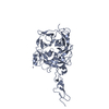
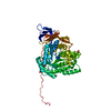
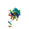
Mass: 54415.508 Da / Num. of mol.: 2 / Fragment: UNP residues 193-667
Source method: isolated from a genetically manipulated source
Details: Expression of selenomethionine labeled Hhip (Se-Met Hhip) was carried out using ESF921 methionine-free medium (Expression Systems). The medium was supplemented with 100 mg/L selenomethionine ...Details: Expression of selenomethionine labeled Hhip (Se-Met Hhip) was carried out using ESF921 methionine-free medium (Expression Systems). The medium was supplemented with 100 mg/L selenomethionine (Sigma Aldrich) 12 and 36 h after virus infection.
Source: (gene. exp.) Homo sapiens (human)
Gene: HHIP, HHIP ORFNames: UNQ5825/PRO19644, HIP, UNQ5825/PRO19644
Plasmid: pENTR/D-TOPO with honeybee melittin secretion signal
Cell line (production host): insect cells / Production host: Trichoplusia ni (cabbage looper) / References: UniProt: Q96QV1

-
Experimental details

-
Experiment

Experiment
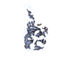
Method: X-RAY DIFFRACTION / Number of used crystals: 1

-
Sample preparation

CrystalDensity Matthews: 4.15 Å3/Da / Density % sol: 70.39 %
Crystal growTemperature: 292 K / Method: vapor diffusion, hanging drop / pH: 7.2
Details: Protein samples (0.3-2 ul) were dehydrated over 500 ul of reservoir solution of 20 mM Hepes pH 7.2 and 3 M NaCl, VAPOR DIFFUSION, HANGING DROP, temperature 292K

-
Data collection

DiffractionMean temperature: 100 K
Diffraction sourceSource: SYNCHROTRON / Site: SSRL / Beamline: BL9-2 / Wavelength: 0.97940, 0.97955, 0.91176
DetectorType: MARMOSAIC 325 mm CCD / Detector: CCD / Date: Feb 13, 2007
Details: Flat collimating mirror, double crystal monochromator, toroid focusing mirror
RadiationMonochromator: Double crystal monochromator Si(111). A second set of Si(220) crystals is also available.
Protocol: MAD / Monochromatic (M) / Laue (L): M / Scattering type: x-ray
Radiation wavelength
IDWavelength (Å)Relative weight
10.97941
20.979551
30.911761
ReflectionResolution: 3.1→30 Å / Num. all: 33725 / Num. obs: 33657 / % possible obs: 99.8 % / Observed criterion σ(F): 0 / Observed criterion σ(I): -3 / Redundancy: 5.3 % / Rsym value: 0.074 / Net I/σ(I): 21.2
Reflection shellResolution: 3.1→3.21 Å / Redundancy: 5.1 % / Mean I/σ(I) obs: 2.9 / Rsym value: 0.568 / % possible all: 99.2

-
Processing

Software
NameVersionClassification
Blu-Icedata collection
SHARPphasing
REFMAC5.2.0019refinement
HKL-2000data reduction
HKL-2000data scaling
RefinementMethod to determine structure: MAD / Resolution: 3.1→30 Å / Cor.coef. Fo:Fc: 0.915 / Cor.coef. Fo:Fc free: 0.893 / SU B: 35.622 / SU ML: 0.29 / TLS residual ADP flag: LIKELY RESIDUAL / Cross valid method: THROUGHOUT / ESU R: 0.842 / ESU R Free: 0.369 / Stereochemistry target values: MAXIMUM LIKELIHOOD / Details: HYDROGENS HAVE BEEN ADDED IN THE RIDING POSITIONS
RfactorNum. reflection% reflectionSelection details
Rfree0.25237 1709 5.1 %RANDOM
Rwork0.21748 ---
obs0.21926 31970 99.94 %-
all-31990 --
Solvent computationIon probe radii: 0.8 Å / Shrinkage radii: 0.8 Å / VDW probe radii: 1.4 Å / Solvent model: MASK
Displacement parametersBiso mean: 72.938 Å2
Baniso -1Baniso -2Baniso -3
1-2.03 Å21.01 Å20 Å2
2--2.03 Å20 Å2
3----3.04 Å2
Refinement stepCycle: LAST / Resolution: 3.1→30 Å
ProteinNucleic acidLigandSolventTotal
Num. atoms6868 0 0 0 6868
Refine LS restraints
Refine-IDTypeDev idealDev ideal targetNumber
X-RAY DIFFRACTIONr_bond_refined_d0.0080.0217036
X-RAY DIFFRACTIONr_bond_other_d
X-RAY DIFFRACTIONr_angle_refined_deg1.2031.9669511
X-RAY DIFFRACTIONr_angle_other_deg
X-RAY DIFFRACTIONr_dihedral_angle_1_deg6.9065872
X-RAY DIFFRACTIONr_dihedral_angle_2_deg33.31523.488324
X-RAY DIFFRACTIONr_dihedral_angle_3_deg20.045151191
X-RAY DIFFRACTIONr_dihedral_angle_4_deg20.5211550
X-RAY DIFFRACTIONr_chiral_restr0.0840.21003
X-RAY DIFFRACTIONr_gen_planes_refined0.0030.025384
X-RAY DIFFRACTIONr_gen_planes_other
X-RAY DIFFRACTIONr_nbd_refined0.2110.22882
X-RAY DIFFRACTIONr_nbd_other
X-RAY DIFFRACTIONr_nbtor_refined0.3090.24552
X-RAY DIFFRACTIONr_nbtor_other
X-RAY DIFFRACTIONr_xyhbond_nbd_refined0.1230.2220
X-RAY DIFFRACTIONr_xyhbond_nbd_other
X-RAY DIFFRACTIONr_metal_ion_refined
X-RAY DIFFRACTIONr_metal_ion_other
X-RAY DIFFRACTIONr_symmetry_vdw_refined0.1690.242
X-RAY DIFFRACTIONr_symmetry_vdw_other
X-RAY DIFFRACTIONr_symmetry_hbond_refined0.1360.24
X-RAY DIFFRACTIONr_symmetry_hbond_other
X-RAY DIFFRACTIONr_symmetry_metal_ion_refined
X-RAY DIFFRACTIONr_symmetry_metal_ion_other
X-RAY DIFFRACTIONr_mcbond_it2.3312.54441
X-RAY DIFFRACTIONr_mcbond_other
X-RAY DIFFRACTIONr_mcangle_it3.91356989
X-RAY DIFFRACTIONr_scbond_it2.1782.52860
X-RAY DIFFRACTIONr_scangle_it3.53152522
X-RAY DIFFRACTIONr_rigid_bond_restr
X-RAY DIFFRACTIONr_sphericity_free
X-RAY DIFFRACTIONr_sphericity_bonded
LS refinement shellResolution: 3.101→3.164 Å / Total num. of bins used: 25
RfactorNum. reflection% reflection
Rfree0.382 105 -
Rwork0.297 1858 -
obs--99.8 %
Refinement TLS params.

Method: refined / Refine-ID: X-RAY DIFFRACTION

IDL112)L122)L132)L222)L232)L332)S11 (Å °)S12 (Å °)S13 (Å °)S21 (Å °)S22 (Å °)S23 (Å °)S31 (Å °)S32 (Å °)S33 (Å °)T112)T122)T132)T222)T232)T332)Origin x (Å)Origin y (Å)Origin z (Å)
13.8249-0.3983-0.37071.83360.01674.16330.00070.87550.1177-0.26250.05620.00340.01160.2507-0.0569-0.3548-0.1444-0.0226-0.0409-0.0466-0.1698-89.207730.764848.6769
218.67263.7166-7.0222.4116-1.25012.6537-0.14870.9052-0.4648-0.4848-0.7128-0.26960.6866-0.0480.8616-0.24910.20070.14520.7166-0.169-0.0718-61.873720.280338.3951
322.4648-2.6355-4.65789.05090.808313.72570.35510.97482.1283-0.3953-0.1893-0.2305-1.3561-0.8369-0.1658-0.27640.03790.1047-0.19820.0823-0.0381-39.212925.904330.3958
43.0675-0.6101-0.51753.29341.69216.9591-0.2244-0.682-0.23140.91870.19420.0581.32680.28020.03020.04560.06660.0588-0.27180.1195-0.2398-93.826825.724898.4159
56.619-6.5176-4.158811.78238.09945.602-0.2671-0.75370.03290.8364-0.73440.69560.5133-1.73161.0016-0.26550.01120.26220.3347-0.2546-0.0139-115.698244.2967110.5126
617.3274-12.4133-3.08317.88945.962814.4959-0.5721-0.66731.4019-0.07510.953-1.5982-0.40951.9245-0.3809-0.2171-0.29630.11240.2653-0.26250.098-121.15963.552125.1252
Refinement TLS group
IDRefine-IDRefine TLS-IDAuth asym-IDAuth seq-ID
1X-RAY DIFFRACTION1A215 - 603
2X-RAY DIFFRACTION2A604 - 637
3X-RAY DIFFRACTION3A638 - 669
4X-RAY DIFFRACTION4B215 - 603
5X-RAY DIFFRACTION5B604 - 637
6X-RAY DIFFRACTION6B638 - 669

+
About Yorodumi

-
News

-
Feb 9, 2022. New format data for meta-information of EMDB entries

New format data for meta-information of EMDB entries

  • Version 3 of the EMDB header file is now the official format.
  • The previous official version 1.9 will be removed from the archive.

Related info.:EMDB header

External links:wwPDB to switch to version 3 of the EMDB data model

-
Aug 12, 2020. Covid-19 info

Covid-19 info

URL: https://pdbj.org/emnavi/covid19.php

New page: Covid-19 featured information page in EM Navigator.

Related info.:Covid-19 info / Mar 5, 2020. Novel coronavirus structure data

+
Mar 5, 2020. Novel coronavirus structure data

Novel coronavirus structure data

Related info.:Yorodumi Speices / Aug 12, 2020. Covid-19 info

External links:COVID-19 featured content - PDBj / Molecule of the Month (242):Coronavirus Proteases

+
Jan 31, 2019. EMDB accession codes are about to change! (news from PDBe EMDB page)

EMDB accession codes are about to change! (news from PDBe EMDB page)

  • The allocation of 4 digits for EMDB accession codes will soon come to an end. Whilst these codes will remain in use, new EMDB accession codes will include an additional digit and will expand incrementally as the available range of codes is exhausted. The current 4-digit format prefixed with “EMD-” (i.e. EMD-XXXX) will advance to a 5-digit format (i.e. EMD-XXXXX), and so on. It is currently estimated that the 4-digit codes will be depleted around Spring 2019, at which point the 5-digit format will come into force.
  • The EM Navigator/Yorodumi systems omit the EMD- prefix.

Related info.:Q: What is EMD? / ID/Accession-code notation in Yorodumi/EM Navigator

External links:EMDB Accession Codes are Changing Soon! / Contact to PDBj

+
Jul 12, 2017. Major update of PDB

Major update of PDB

  • wwPDB released updated PDB data conforming to the new PDBx/mmCIF dictionary.
  • This is a major update changing the version number from 4 to 5, and with Remediation, in which all the entries are updated.
  • In this update, many items about electron microscopy experimental information are reorganized (e.g. em_software).
  • Now, EM Navigator and Yorodumi are based on the updated data.

External links:wwPDB Remediation / Enriched Model Files Conforming to OneDep Data Standards Now Available in the PDB FTP Archive

-
Yorodumi

Thousand views of thousand structures

  • Yorodumi is a browser for structure data from EMDB, PDB, SASBDB, etc.
  • This page is also the successor to EM Navigator detail page, and also detail information page/front-end page for Omokage search.
  • The word "yorodu" (or yorozu) is an old Japanese word meaning "ten thousand". "mi" (miru) is to see.

Related info.:EMDB / PDB / SASBDB / Comparison of 3 databanks / Yorodumi Search / Aug 31, 2016. New EM Navigator & Yorodumi / Yorodumi Papers / Jmol/JSmol / Function and homology information / Changes in new EM Navigator and Yorodumi

Read more