 Movie
Movie Controller


+ Open data

- Basic information
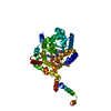
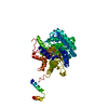
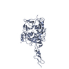
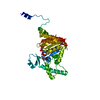
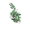
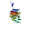
| Entry | Database: PDB / ID: 3ho3 | ||||||
|---|---|---|---|---|---|---|---|
| Title | Crystal structure of Hedgehog-interacting protein (HHIP) | ||||||
 Components Components | Hedgehog-interacting protein | ||||||
 Keywords Keywords | SIGNALING PROTEIN / receptor ectodomain / six-bladed-propeller domain / EGF domain / disulfide bond / Cell membrane / EGF-like domain / Glycoprotein / Membrane / Secreted | ||||||
| Function / homology |  Function and homology information Function and homology informationregulation of fibroblast growth factor receptor signaling pathway / hedgehog family protein binding / Ligand-receptor interactions / dorsal/ventral pattern formation / skeletal system morphogenesis / ciliary membrane / epithelial tube branching involved in lung morphogenesis / neuroblast proliferation / negative regulation of signal transduction / negative regulation of smoothened signaling pathway ...regulation of fibroblast growth factor receptor signaling pathway / hedgehog family protein binding / Ligand-receptor interactions / dorsal/ventral pattern formation / skeletal system morphogenesis / ciliary membrane / epithelial tube branching involved in lung morphogenesis / neuroblast proliferation / negative regulation of signal transduction / negative regulation of smoothened signaling pathway / cell surface / signal transduction / extracellular region / zinc ion binding / plasma membrane / cytoplasm Similarity search - Function | ||||||
| Biological species |  Homo sapiens (human) Homo sapiens (human) | ||||||
| Method |  X-RAY DIFFRACTION / SYNCHROTRON / MOLECULAR REPLACEMENT / Resolution: 2.9 Å X-RAY DIFFRACTION / SYNCHROTRON / MOLECULAR REPLACEMENT / Resolution: 2.9 Å | ||||||
 Authors Authors | Bosanac, I. / Hymowitz, S.G. | ||||||
 Citation Citation | Journal: Nat.Struct.Mol.Biol. / Year: 2009 Title: The structure of SHH in complex with HHIP reveals a recognition role for the Shh pseudo active site in signaling. Authors: Bosanac, I. / Maun, H.R. / Scales, S.J. / Wen, X. / Lingel, A. / Bazan, J.F. / de Sauvage, F.J. / Hymowitz, S.G. / Lazarus, R.A. | ||||||
| History |
|
- Structure visualization
| Structure viewer | Molecule: MolmilJmol/JSmol |
|---|
- Downloads & links
-Download
| PDBx/mmCIF format | 3ho3.cif.gz | 100 KB | Display | PDBx/mmCIF format |
|---|---|---|---|---|
| PDB format | pdb3ho3.ent.gz | 76.1 KB | Display | PDB format |
| PDBx/mmJSON format | 3ho3.json.gz | Tree view | PDBx/mmJSON format | |
| Others |  Other downloads Other downloads |
-Validation report
| Arichive directory | https://data.pdbj.org/pub/pdb/validation_reports/ho/3ho3ftp://data.pdbj.org/pub/pdb/validation_reports/ho/3ho3 | HTTPS FTP |
|---|
-Related structure data
| Related structure data |  3ho4SC  3ho5C S: Starting model for refinement C: citing same article ( |
|---|---|
| Similar structure data |
-Links
 PDBj
PDBj


- Assembly
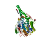
| Deposited unit | 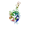
| ||||||||
|---|---|---|---|---|---|---|---|---|---|
| 1 |
| ||||||||
| Unit cell |
|
-Components
| #1: Protein | Mass: 54087.246 Da / Num. of mol.: 1 / Fragment: UNP residues 193-667 Source method: isolated from a genetically manipulated source Source: (gene. exp.) Homo sapiens (human)Gene: HHIP, HHIP ORFNames: UNQ5825/PRO19644, HIP, UNQ5825/PRO19644 Plasmid: pENTR/D-TOPO with honeybee melittin secretion signal Cell line (production host): insect cells / Production host:  Trichoplusia ni (cabbage looper) / References: UniProt: Q96QV1 Trichoplusia ni (cabbage looper) / References: UniProt: Q96QV1 |
|---|---|
| Has protein modification | Y |
-Experimental details
-Experiment
| Experiment | Method: X-RAY DIFFRACTION / Number of used crystals: 1 |
|---|
- Sample preparation
| Crystal | Density Matthews: 2.97 Å3/Da / Density % sol: 58.61 % |
|---|---|
| Crystal grow | Temperature: 292 K / Method: vapor diffusion, hanging drop / pH: 5.6 Details: 0.1 M sodium acetate, 0.2 M ammonium sulfate, 22-24% PEG 4000 and 1-2% dioxane, pH 5.6, VAPOR DIFFUSION, HANGING DROP, temperature 292K |
-Data collection
| Diffraction | Mean temperature: 100 K |
|---|---|
| Diffraction source | Source: SYNCHROTRON / Site: ALS  / Beamline: 5.0.2 / Wavelength: 1 Å / Beamline: 5.0.2 / Wavelength: 1 Å |
| Detector | Type: ADSC QUANTUM 315 / Detector: CCD / Date: Aug 26, 2006 Details: vertically collimating premirror, LN2 cooled double-crystal silicon (111) monochromator, toroidal focusing M2 mirror |
| Radiation | Monochromator: LN2 cooled double-crystal silicon (111) monochromator with toroidal focusing M2 mirror Protocol: SINGLE WAVELENGTH / Monochromatic (M) / Laue (L): M / Scattering type: x-ray |
| Radiation wavelength | Wavelength: 1 Å / Relative weight: 1 |
| Reflection | Resolution: 2.8→30 Å / Num. all: 14785 / Num. obs: 14755 / % possible obs: 99.8 % / Observed criterion σ(I): -3 / Redundancy: 4.7 % / Rsym value: 0.089 / Net I/σ(I): 14.4 |
| Reflection shell | Resolution: 2.8→2.9 Å / Redundancy: 4.8 % / Mean I/σ(I) obs: 3.5 / Rsym value: 0.491 / % possible all: 100 |
- Processing
| Software |
| ||||||||||||||||||||||||||||||||||||||||||||||||||||||||||||||||||||||||||||||||||||||||||||||||||||
|---|---|---|---|---|---|---|---|---|---|---|---|---|---|---|---|---|---|---|---|---|---|---|---|---|---|---|---|---|---|---|---|---|---|---|---|---|---|---|---|---|---|---|---|---|---|---|---|---|---|---|---|---|---|---|---|---|---|---|---|---|---|---|---|---|---|---|---|---|---|---|---|---|---|---|---|---|---|---|---|---|---|---|---|---|---|---|---|---|---|---|---|---|---|---|---|---|---|---|---|---|---|
| Refinement | Method to determine structure: MOLECULAR REPLACEMENT Starting model: PDB entry 3HO4 Resolution: 2.9→30 Å / Cor.coef. Fo:Fc: 0.932 / Cor.coef. Fo:Fc free: 0.912 / SU B: 45.17 / SU ML: 0.386 / TLS residual ADP flag: LIKELY RESIDUAL / Cross valid method: THROUGHOUT / σ(F): 0 / σ(I): -3 / ESU R: 4.696 / ESU R Free: 0.43 / Stereochemistry target values: MAXIMUM LIKELIHOOD / Details: HYDROGENS HAVE BEEN ADDED IN THE RIDING POSITIONS
| ||||||||||||||||||||||||||||||||||||||||||||||||||||||||||||||||||||||||||||||||||||||||||||||||||||
| Solvent computation | Ion probe radii: 0.8 Å / Shrinkage radii: 0.8 Å / VDW probe radii: 1.4 Å / Solvent model: MASK | ||||||||||||||||||||||||||||||||||||||||||||||||||||||||||||||||||||||||||||||||||||||||||||||||||||
| Displacement parameters | Biso mean: 94.581 Å2
| ||||||||||||||||||||||||||||||||||||||||||||||||||||||||||||||||||||||||||||||||||||||||||||||||||||
| Refinement step | Cycle: LAST / Resolution: 2.9→30 Å
| ||||||||||||||||||||||||||||||||||||||||||||||||||||||||||||||||||||||||||||||||||||||||||||||||||||
| Refine LS restraints |
| ||||||||||||||||||||||||||||||||||||||||||||||||||||||||||||||||||||||||||||||||||||||||||||||||||||
| LS refinement shell | Resolution: 2.9→2.959 Å / Total num. of bins used: 25
| ||||||||||||||||||||||||||||||||||||||||||||||||||||||||||||||||||||||||||||||||||||||||||||||||||||
| Refinement TLS params. | Method: refined / Refine-ID: X-RAY DIFFRACTION
| ||||||||||||||||||||||||||||||||||||||||||||||||||||||||||||||||||||||||||||||||||||||||||||||||||||
| Refinement TLS group |
|
