 Movie
Movie Controller

[English] 日本語
 Yorodumi
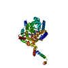
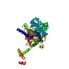
- PDB-1tj2: Crystal structure of E. coli PutA proline dehydrogenase domain (r... -
+ Open data

- Basic information
| Entry | Database: PDB / ID: 1tj2 | ||||||
|---|---|---|---|---|---|---|---|
| Title | Crystal structure of E. coli PutA proline dehydrogenase domain (residues 86-669) complexed with acetate | ||||||
 Components Components | Bifunctional putA protein | ||||||
 Keywords Keywords | OXIDOREDUCTASE / beta/alpha barrel / flavoenzyme / FAD / proline catabolism | ||||||
| Function / homology |  Function and homology information Function and homology informationproline dehydrogenase / proline dehydrogenase activity / L-glutamate gamma-semialdehyde dehydrogenase / L-glutamate gamma-semialdehyde dehydrogenase activity / : / L-proline biosynthetic process / DNA-binding transcription repressor activity / cis-regulatory region sequence-specific DNA binding / cytoplasmic side of plasma membrane / flavin adenine dinucleotide binding ...proline dehydrogenase / proline dehydrogenase activity / L-glutamate gamma-semialdehyde dehydrogenase / L-glutamate gamma-semialdehyde dehydrogenase activity / : / L-proline biosynthetic process / DNA-binding transcription repressor activity / cis-regulatory region sequence-specific DNA binding / cytoplasmic side of plasma membrane / flavin adenine dinucleotide binding / response to oxidative stress / sequence-specific DNA binding / negative regulation of DNA-templated transcription / DNA-templated transcription / protein homodimerization activity / DNA binding / identical protein binding / plasma membrane / cytosol Similarity search - Function | ||||||
| Biological species |  | ||||||
| Method |  X-RAY DIFFRACTION / SYNCHROTRON / MOLECULAR REPLACEMENT / Resolution: 2.05 Å X-RAY DIFFRACTION / SYNCHROTRON / MOLECULAR REPLACEMENT / Resolution: 2.05 Å | ||||||
 Authors Authors | Tanner, J.J. / Zhang, M. / White, T.A. / Schuermann, J.P. / Baban, B.A. / Becker, D.F. | ||||||
 Citation Citation | Journal: Biochemistry / Year: 2004 Title: Structures of the Escherichia coli PutA proline dehydrogenase domain in complex with competitive inhibitors Authors: Zhang, M. / White, T.A. / Schuermann, J.P. / Baban, B.A. / Becker, D.F. / Tanner, J.J. #1: Journal: Acta Crystallogr.,Sect.D / Year: 2004Title: Detection of L-lactate in polyethylene glycol solutions confirms the identity of the active-site ligand in a proline dehydrogenase structure Authors: Zhang, M. / Tanner, J.J. | ||||||
| History |
|
- Structure visualization
| Structure viewer | Molecule: MolmilJmol/JSmol |
|---|
- Downloads & links
-Download
| PDBx/mmCIF format | 1tj2.cif.gz | 110.7 KB | Display | PDBx/mmCIF format |
|---|---|---|---|---|
| PDB format | pdb1tj2.ent.gz | 81 KB | Display | PDB format |
| PDBx/mmJSON format | 1tj2.json.gz | Tree view | PDBx/mmJSON format | |
| Others |  Other downloads Other downloads |
-Validation report
| Arichive directory | https://data.pdbj.org/pub/pdb/validation_reports/tj/1tj2ftp://data.pdbj.org/pub/pdb/validation_reports/tj/1tj2 | HTTPS FTP |
|---|
-Related structure data
| Related structure data |  1tiwC  1tj0C  1tj1C  1k87 C: citing same article ( S: Starting model for refinement |
|---|---|
| Similar structure data |


-Links
 PDBj
PDBj

- Assembly
| Deposited unit | 
| ||||||||
|---|---|---|---|---|---|---|---|---|---|
| 1 |
| ||||||||
| 2 | 
| ||||||||
| Unit cell |
|
-Components
| #1: Protein | Mass: 64665.871 Da / Num. of mol.: 1 Fragment: E. coli PutA proline dehydrogenase domain (residues 86-669) Source method: isolated from a genetically manipulated source Source: (gene. exp.) |
|---|---|
| #2: Chemical | ChemComp-ACT /   Mass: 59.044 Da / Num. of mol.: 1 / Source method: obtained synthetically / Formula: C2H3O2 Mass: 59.044 Da / Num. of mol.: 1 / Source method: obtained synthetically / Formula: C2H3O2 |
| #3: Chemical | ChemComp-FAD /   Mass: 785.550 Da / Num. of mol.: 1 / Source method: obtained synthetically / Formula: C27H33N9O15P2 / Comment: FAD*YM Mass: 785.550 Da / Num. of mol.: 1 / Source method: obtained synthetically / Formula: C27H33N9O15P2 / Comment: FAD*YM |
| #4: Water | ChemComp-HOH /  Mass: 18.015 Da / Num. of mol.: 193 / Source method: isolated from a natural source / Formula: H2O Mass: 18.015 Da / Num. of mol.: 193 / Source method: isolated from a natural source / Formula: H2O |
-Experimental details
-Experiment
| Experiment | Method: X-RAY DIFFRACTION / Number of used crystals: 1 |
|---|
- Sample preparation
| Crystal | Density Matthews: 2.9 Å3/Da / Density % sol: 58 % |
|---|---|
| Crystal grow | Temperature: 295 K / Method: vapor diffusion, sitting drop / pH: 5.7 Details: 13-15 % PEG 3350, 60-190 mM citrate buffer, pH 5.7, VAPOR DIFFUSION, SITTING DROP, temperature 295K |
-Data collection
| Diffraction | Mean temperature: 173 K |
|---|---|
| Diffraction source | Source: SYNCHROTRON / Site: APS  / Beamline: 19-ID / Wavelength: 0.97856 Å / Beamline: 19-ID / Wavelength: 0.97856 Å |
| Detector | Type: CUSTOM-MADE / Detector: CCD / Date: Nov 15, 2003 / Details: APS 19ID |
| Radiation | Monochromator: APS 19ID / Protocol: SINGLE WAVELENGTH / Monochromatic (M) / Laue (L): M / Scattering type: x-ray |
| Radiation wavelength | Wavelength: 0.97856 Å / Relative weight: 1 |
| Reflection | Resolution: 2.05→99 Å / Num. all: 46790 / Num. obs: 45470 / % possible obs: 97 % / Observed criterion σ(F): 0 / Observed criterion σ(I): 0 / Redundancy: 6.8 % / Biso Wilson estimate: 40 Å2 / Rmerge(I) obs: 0.084 / Rsym value: 0.084 / Net I/σ(I): 14 |
| Reflection shell | Resolution: 2.05→2.12 Å / Redundancy: 5.4 % / Rmerge(I) obs: 0.405 / Mean I/σ(I) obs: 2.9 / Num. unique all: 4232 / Rsym value: 0.405 / % possible all: 91 |
- Processing
| Software |
| ||||||||||||||||||||||||||||||||||||||||||||||||||||||||||||||||||||||||||||||||||||||||||||||||||||
|---|---|---|---|---|---|---|---|---|---|---|---|---|---|---|---|---|---|---|---|---|---|---|---|---|---|---|---|---|---|---|---|---|---|---|---|---|---|---|---|---|---|---|---|---|---|---|---|---|---|---|---|---|---|---|---|---|---|---|---|---|---|---|---|---|---|---|---|---|---|---|---|---|---|---|---|---|---|---|---|---|---|---|---|---|---|---|---|---|---|---|---|---|---|---|---|---|---|---|---|---|---|
| Refinement | Method to determine structure: MOLECULAR REPLACEMENT Starting model: PDB ENTRY 1k87 1k87 Resolution: 2.05→22.7 Å / Cor.coef. Fo:Fc: 0.943 / Cor.coef. Fo:Fc free: 0.914 / SU B: 3.693 / SU ML: 0.101 / TLS residual ADP flag: LIKELY RESIDUAL / Cross valid method: THROUGHOUT / σ(F): 0 / σ(I): 0 / ESU R: 0.158 / ESU R Free: 0.153 / Stereochemistry target values: MAXIMUM LIKELIHOOD / Details: HYDROGENS HAVE BEEN ADDED IN THE RIDING POSITIONS
| ||||||||||||||||||||||||||||||||||||||||||||||||||||||||||||||||||||||||||||||||||||||||||||||||||||
| Solvent computation | Ion probe radii: 0.8 Å / Shrinkage radii: 0.8 Å / VDW probe radii: 1.4 Å / Solvent model: BABINET MODEL WITH MASK | ||||||||||||||||||||||||||||||||||||||||||||||||||||||||||||||||||||||||||||||||||||||||||||||||||||
| Displacement parameters | Biso mean: 27.081 Å2
| ||||||||||||||||||||||||||||||||||||||||||||||||||||||||||||||||||||||||||||||||||||||||||||||||||||
| Refine analyze |
| ||||||||||||||||||||||||||||||||||||||||||||||||||||||||||||||||||||||||||||||||||||||||||||||||||||
| Refinement step | Cycle: LAST / Resolution: 2.05→22.7 Å
| ||||||||||||||||||||||||||||||||||||||||||||||||||||||||||||||||||||||||||||||||||||||||||||||||||||
| Refine LS restraints |
| ||||||||||||||||||||||||||||||||||||||||||||||||||||||||||||||||||||||||||||||||||||||||||||||||||||
| LS refinement shell | Resolution: 2.05→2.103 Å / Total num. of bins used: 20
| ||||||||||||||||||||||||||||||||||||||||||||||||||||||||||||||||||||||||||||||||||||||||||||||||||||
| Refinement TLS params. | Method: refined / Origin x: 6.827 Å / Origin y: 45.312 Å / Origin z: 52.439 Å
| ||||||||||||||||||||||||||||||||||||||||||||||||||||||||||||||||||||||||||||||||||||||||||||||||||||
| Refinement TLS group |
|
