Protocol: SINGLE WAVELENGTH / Monochromatic (M) / Laue (L): M / Scattering type: x-ray
Radiation wavelength
Wavelength: 0.979 Å / Relative weight: 1
Reflection
Resolution: 1.48→40 Å / Num. obs: 41183 / % possible obs: 94.3 % / Redundancy: 4.4 % / Rmerge(I) obs: 0.081 / Χ2: 1.302 / Net I/σ(I): 15.4
Reflection shell
Resolution (Å)
Redundancy (%)
Rmerge(I) obs
Num. unique all
Χ2
% possible all
1.48-1.53
2.2
0.334
5136
0.936
62
1.53-1.59
2.3
0.261
7058
0.961
83.7
1.59-1.67
2.7
0.26
8030
0.951
97.4
1.67-1.75
3.2
0.262
8362
0.951
100
1.75-1.86
3.7
0.253
8350
0.966
100
1.86-2.01
4.6
0.262
8301
1.189
100
2.01-2.21
5.8
0.227
8343
1.03
100
2.21-2.53
6.3
0.164
8363
1.323
100
2.53-3.19
6.2
0.083
8318
1.663
100
3.19-50
5.7
0.045
8354
1.848
99.8
-
Processing
Software
Name
Version
Classification
NB
DENZO
datareduction
SCALEPACK
datascaling
REFMAC
5.2.0005
refinement
PDB_EXTRACT
2
dataextraction
Refinement
Method to determine structure: SAD / Resolution: 1.5→40 Å / Cor.coef. Fo:Fc: 0.975 / Cor.coef. Fo:Fc free: 0.955 / SU B: 2.848 / SU ML: 0.048 / Cross valid method: THROUGHOUT / σ(F): 0 / ESU R: 0.072 / ESU R Free: 0.069 / Stereochemistry target values: MAXIMUM LIKELIHOOD / Details: HYDROGENS HAVE BEEN ADDED IN THE RIDING POSITIONS
Rfactor
Num. reflection
% reflection
Selection details
Rfree
0.203
2063
5 %
RANDOM
Rwork
0.156
-
-
-
all
0.158
-
-
-
obs
0.157
41134
97.72 %
-
Solvent computation
Ion probe radii: 0.8 Å / Shrinkage radii: 0.8 Å / VDW probe radii: 1.2 Å / Solvent model: MASK
Displacement parameters
Biso mean: 21.551 Å2
Baniso -1
Baniso -2
Baniso -3
1-
0.6 Å2
0.3 Å2
0 Å2
2-
-
0.6 Å2
0 Å2
3-
-
-
-0.89 Å2
Refinement step
Cycle: LAST / Resolution: 1.5→40 Å
Protein
Nucleic acid
Ligand
Solvent
Total
Num. atoms
1592
0
10
323
1925
Refine LS restraints
Refine-ID
Type
Dev ideal
Dev ideal target
Number
X-RAY DIFFRACTION
r_bond_refined_d
0.02
0.022
1626
X-RAY DIFFRACTION
r_bond_other_d
0.003
0.02
1468
X-RAY DIFFRACTION
r_angle_refined_deg
1.69
1.983
2197
X-RAY DIFFRACTION
r_angle_other_deg
0.898
3
3435
X-RAY DIFFRACTION
r_dihedral_angle_1_deg
6.102
5
199
X-RAY DIFFRACTION
r_dihedral_angle_2_deg
34.71
24.868
76
X-RAY DIFFRACTION
r_dihedral_angle_3_deg
13.119
15
302
X-RAY DIFFRACTION
r_dihedral_angle_4_deg
18.893
15
10
X-RAY DIFFRACTION
r_chiral_restr
0.141
0.2
246
X-RAY DIFFRACTION
r_gen_planes_refined
0.008
0.02
1785
X-RAY DIFFRACTION
r_gen_planes_other
0.001
0.02
308
X-RAY DIFFRACTION
r_nbd_refined
0.222
0.2
354
X-RAY DIFFRACTION
r_nbd_other
0.204
0.2
1566
X-RAY DIFFRACTION
r_nbtor_refined
0.187
0.2
833
X-RAY DIFFRACTION
r_nbtor_other
0.089
0.2
958
X-RAY DIFFRACTION
r_xyhbond_nbd_refined
0.178
0.2
226
X-RAY DIFFRACTION
r_symmetry_vdw_refined
0.461
0.2
13
X-RAY DIFFRACTION
r_symmetry_vdw_other
0.181
0.2
44
X-RAY DIFFRACTION
r_symmetry_hbond_refined
0.238
0.2
33
X-RAY DIFFRACTION
r_mcbond_it
2.236
1.5
1280
X-RAY DIFFRACTION
r_mcbond_other
1.208
1.5
405
X-RAY DIFFRACTION
r_mcangle_it
2.583
2
1613
X-RAY DIFFRACTION
r_scbond_it
4.566
3
722
X-RAY DIFFRACTION
r_scangle_it
5.656
4.5
584
LS refinement shell
Resolution: 1.5→1.539 Å / Total num. of bins used: 20
Rfactor
Num. reflection
% reflection
Rfree
0.239
123
-
Rwork
0.176
2274
-
obs
-
2397
78.8 %
Refinement TLS params.
Method: refined / Refine-ID: X-RAY DIFFRACTION
ID
L11 (°2)
L12 (°2)
L13 (°2)
L22 (°2)
L23 (°2)
L33 (°2)
S11 (Å °)
S12 (Å °)
S13 (Å °)
S21 (Å °)
S22 (Å °)
S23 (Å °)
S31 (Å °)
S32 (Å °)
S33 (Å °)
T11 (Å2)
T12 (Å2)
T13 (Å2)
T22 (Å2)
T23 (Å2)
T33 (Å2)
Origin x (Å)
Origin y (Å)
Origin z (Å)
1
1.6379
-0.5147
0.7637
0.8656
-0.1643
3.5562
0.0546
-0.2197
0.1051
0.0155
-0.0752
0.0208
-0.2072
-0.2202
0.0206
-0.0745
-0.0624
0.0084
0.102
-0.0144
-0.028
24.571
24.616
38.653
2
0.4556
-0.1468
1.0678
0.2936
-0.5799
3.8328
0.1318
0.0178
0.0094
0.0029
-0.154
-0.0026
0.0794
0.2357
0.0222
-0.0761
0.0203
0.0061
0.0571
-0.0073
-0.0373
26.128
22.106
14.23
Refinement TLS group
Refine-ID: X-RAY DIFFRACTION / Selection: ALL / Auth asym-ID: A / Label asym-ID: A
ID
Refine TLS-ID
Auth seq-ID
Label seq-ID
1
1
391 - 458
1 - 68
2
2
459 - 590
69 - 200
+
About Yorodumi
-
News
-
Feb 9, 2022. New format data for meta-information of EMDB entries
New format data for meta-information of EMDB entries
Version 3 of the EMDB header file is now the official format.
The previous official version 1.9 will be removed from the archive.
In the structure databanks used in Yorodumi, some data are registered as the other names, "COVID-19 virus" and "2019-nCoV". Here are the details of the virus and the list of structure data.
Jan 31, 2019. EMDB accession codes are about to change! (news from PDBe EMDB page)
EMDB accession codes are about to change! (news from PDBe EMDB page)
The allocation of 4 digits for EMDB accession codes will soon come to an end. Whilst these codes will remain in use, new EMDB accession codes will include an additional digit and will expand incrementally as the available range of codes is exhausted. The current 4-digit format prefixed with “EMD-” (i.e. EMD-XXXX) will advance to a 5-digit format (i.e. EMD-XXXXX), and so on. It is currently estimated that the 4-digit codes will be depleted around Spring 2019, at which point the 5-digit format will come into force.
The EM Navigator/Yorodumi systems omit the EMD- prefix.
Related info.:Q: What is EMD? / ID/Accession-code notation in Yorodumi/EM Navigator
Yorodumi is a browser for structure data from EMDB, PDB, SASBDB, etc.
This page is also the successor to EM Navigator detail page, and also detail information page/front-end page for Omokage search.
The word "yorodu" (or yorozu) is an old Japanese word meaning "ten thousand". "mi" (miru) is to see.
Related info.:EMDB / PDB / SASBDB / Comparison of 3 databanks / Yorodumi Search / Aug 31, 2016. New EM Navigator & Yorodumi / Yorodumi Papers / Jmol/JSmol / Function and homology information / Changes in new EM Navigator and Yorodumi
 Movie
Movie Controller


 Open data
 Basic information
 Components
 Keywords
 Function and homology information
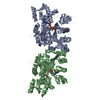
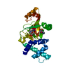
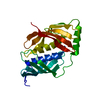
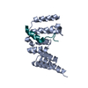
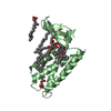
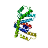
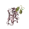
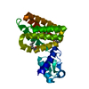
 Salmonella typhimurium (bacteria)
 X-RAY DIFFRACTION /
  Authors
 Citation
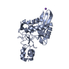
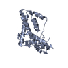
 Structure visualization
 Downloads & links
 Other downloads
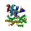


 PDBj
PDBj Assembly
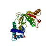

 Mass: 96.063 Da / Num. of mol.: 2 / Source method: obtained synthetically / Formula: SO4
 Mass: 18.015 Da / Num. of mol.: 323 / Source method: isolated from a natural source / Formula: H2O
 Sample preparation
 / Beamline: X9A / Wavelength: 0.979 Å
 Processing