Mass: 18.015 Da / Num. of mol.: 202 / Source method: isolated from a natural source / Formula: H2O
Has protein modification
Y
Sequence details
THIS CONSTRUCT WAS EXPRESSED WITH A PURIFICATION TAG MGSDKIHHHHHHENLYFQG. THE TAG WAS REMOVED WITH ...THIS CONSTRUCT WAS EXPRESSED WITH A PURIFICATION TAG MGSDKIHHHHHHENLYFQG. THE TAG WAS REMOVED WITH TEV PROTEASE LEAVING ONLY A GLYCINE (0) FOLLOWED BY THE TARGET SEQUENCE.
-
Experimental details
-
Experiment
Experiment
Method: X-RAY DIFFRACTION / Number of used crystals: 1
-
Sample preparation
Crystal
Density Matthews: 2.23 Å3/Da / Density % sol: 44.93 %
Crystal grow
Temperature: 277 K / Method: vapor diffusion, sitting drop / pH: 8.17 Details: 24.0000% polyethylene glycol 6000, 1.0000M lithium chloride, 0.1M TRIS pH 8.17, NANODROP, VAPOR DIFFUSION, SITTING DROP, temperature 277K
Monochromator: Single crystal Si(111) bent monochromator (horizontal focusing) Protocol: MAD / Monochromatic (M) / Laue (L): M / Scattering type: x-ray
Radiation wavelength
ID
Wavelength (Å)
Relative weight
1
0.91837
1
2
0.97862
1
3
0.97799
1
Reflection
Resolution: 1.92→29.361 Å / Num. obs: 32709 / % possible obs: 99.6 % / Redundancy: 2.7 % / Biso Wilson estimate: 34.581 Å2 / Rmerge(I) obs: 0.052 / Rsym value: 0.052 / Net I/σ(I): 12.1
Reflection shell
Diffraction-ID: 1
Resolution (Å)
Redundancy (%)
Rmerge(I) obs
Mean I/σ(I) obs
Num. measured all
Num. unique all
Rsym value
% possible all
1.92-1.97
2.7
0.556
1.4
6558
2416
0.556
100
1.97-2.02
2.7
0.415
1.8
6508
2375
0.415
100
2.02-2.08
2.7
0.297
2.5
6266
2290
0.297
100
2.08-2.15
2.7
0.221
3.4
6012
2192
0.221
100
2.15-2.22
2.7
0.167
4.5
5985
2183
0.167
100
2.22-2.29
2.7
0.134
5.5
5828
2129
0.134
100
2.29-2.38
2.7
0.11
6.7
5472
1990
0.11
100
2.38-2.48
2.7
0.093
7.9
5329
1942
0.093
100
2.48-2.59
2.8
0.082
8.7
5186
1882
0.082
100
2.59-2.72
2.8
0.079
8.4
4859
1765
0.079
100
2.72-2.86
2.7
0.076
8.4
4693
1708
0.076
99.9
2.86-3.04
2.7
0.064
8.7
4428
1620
0.064
99.9
3.04-3.25
2.7
0.054
10.6
4092
1490
0.054
99.9
3.25-3.51
2.8
0.046
12.8
3845
1397
0.046
99.7
3.51-3.84
2.8
0.04
14.5
3578
1290
0.04
99.7
3.84-4.29
2.8
0.033
18.8
3247
1174
0.033
99.6
4.29-4.96
2.8
0.032
19.1
2877
1039
0.032
99.4
4.96-6.07
2.7
0.039
15.8
2404
876
0.039
98.9
6.07-8.59
2.7
0.039
16.2
1790
657
0.039
97.9
8.59-29.36
2.4
0.041
15.4
709
294
0.041
74.7
-
Phasing
Phasing
Method: MAD
-
Processing
Software
Name
Version
Classification
NB
REFMAC
5.5.0053
refinement
PHENIX
refinement
SHELX
phasing
MolProbity
3beta29
modelbuilding
SCALA
3.2.5
datascaling
PDB_EXTRACT
3.006
dataextraction
MOSFLM
datareduction
SHELXD
phasing
autoSHARP
phasing
Refinement
Method to determine structure: MAD / Resolution: 1.92→29.361 Å / Cor.coef. Fo:Fc: 0.959 / Cor.coef. Fo:Fc free: 0.941 / Occupancy max: 1 / Occupancy min: 0.3 / SU B: 10.003 / SU ML: 0.128 / TLS residual ADP flag: LIKELY RESIDUAL / Cross valid method: THROUGHOUT / σ(F): 0 / ESU R: 0.172 / ESU R Free: 0.154 Stereochemistry target values: MAXIMUM LIKELIHOOD WITH PHASES Details: 1. HYDROGENS HAVE BEEN ADDED IN THE RIDING POSITIONS. 2. ATOM RECORDS CONTAIN RESIDUAL B FACTORS ONLY. 3. A MET-INHIBITION PROTOCOL WAS USED FOR SELENOMETHIONINE INCORPORATION DURING PROTEIN ...Details: 1. HYDROGENS HAVE BEEN ADDED IN THE RIDING POSITIONS. 2. ATOM RECORDS CONTAIN RESIDUAL B FACTORS ONLY. 3. A MET-INHIBITION PROTOCOL WAS USED FOR SELENOMETHIONINE INCORPORATION DURING PROTEIN EXPRESSION. THE OCCUPANCY OF THE SE ATOMS IN THE MSE RESIDUES WAS REDUCED TO 0.75 FOR THE REDUCED SCATTERING POWER DUE TO PARTIAL S-MET INCORPORATION. 4. CHLORIDE ION AND GLYCEROL MOLECULE FROM THE CRYSTALLIZATION/CRYOPROTECTION SOLUTIONS ARE MODELED.
Rfactor
Num. reflection
% reflection
Selection details
Rfree
0.237
1662
5.1 %
RANDOM
Rwork
0.199
-
-
-
obs
0.201
32709
99.45 %
-
Solvent computation
Ion probe radii: 0.8 Å / Shrinkage radii: 0.8 Å / VDW probe radii: 1.4 Å / Solvent model: BABINET MODEL WITH MASK
In the structure databanks used in Yorodumi, some data are registered as the other names, "COVID-19 virus" and "2019-nCoV". Here are the details of the virus and the list of structure data.
Jan 31, 2019. EMDB accession codes are about to change! (news from PDBe EMDB page)
EMDB accession codes are about to change! (news from PDBe EMDB page)
The allocation of 4 digits for EMDB accession codes will soon come to an end. Whilst these codes will remain in use, new EMDB accession codes will include an additional digit and will expand incrementally as the available range of codes is exhausted. The current 4-digit format prefixed with “EMD-” (i.e. EMD-XXXX) will advance to a 5-digit format (i.e. EMD-XXXXX), and so on. It is currently estimated that the 4-digit codes will be depleted around Spring 2019, at which point the 5-digit format will come into force.
The EM Navigator/Yorodumi systems omit the EMD- prefix.
Related info.:Q: What is EMD? / ID/Accession-code notation in Yorodumi/EM Navigator
Yorodumi is a browser for structure data from EMDB, PDB, SASBDB, etc.
This page is also the successor to EM Navigator detail page, and also detail information page/front-end page for Omokage search.
The word "yorodu" (or yorozu) is an old Japanese word meaning "ten thousand". "mi" (miru) is to see.
Related info.:EMDB / PDB / SASBDB / Comparison of 3 databanks / Yorodumi Search / Aug 31, 2016. New EM Navigator & Yorodumi / Yorodumi Papers / Jmol/JSmol / Function and homology information / Changes in new EM Navigator and Yorodumi
 Movie
Movie Controller

 Yorodumi
 Open data
 Basic information
 Components
 Keywords
 Function and homology information
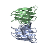
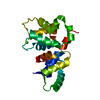
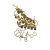
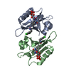
 Ralstonia eutropha (bacteria)
 X-RAY DIFFRACTION /
  Authors
 Citation
 Structure visualization
 Downloads & links
 Other downloads


 PDBj
PDBj Assembly


 Mass: 35.453 Da / Num. of mol.: 2 / Source method: obtained synthetically / Formula: Cl
 Mass: 92.094 Da / Num. of mol.: 1 / Source method: obtained synthetically / Formula: C3H8O3
 Mass: 18.015 Da / Num. of mol.: 202 / Source method: isolated from a natural source / Formula: H2O
 Sample preparation
 / Beamline: BL11-1 / Wavelength: 0.91837,0.97862,0.97799
 Processing