 Movie
Movie Controller


+ Open data

- Basic information
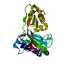
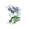
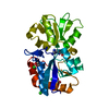
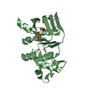
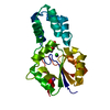
| Entry | Database: PDB / ID: 4nxg | ||||||
|---|---|---|---|---|---|---|---|
| Title | Crystal structure of iLOV-I486z(2LT) at pH 9.0 | ||||||
 Components Components | Phototropin-2 | ||||||
 Keywords Keywords | FLAVOPROTEIN / FLUORESCENT PROTEIN / FMN binding | ||||||
| Function / homology |  Function and homology information Function and homology informationchloroplast relocation / phototropism / blue light signaling pathway / stomatal movement / blue light photoreceptor activity / response to blue light / plastid / circadian rhythm / kinase activity / FMN binding ...chloroplast relocation / phototropism / blue light signaling pathway / stomatal movement / blue light photoreceptor activity / response to blue light / plastid / circadian rhythm / kinase activity / FMN binding / non-specific serine/threonine protein kinase / protein serine kinase activity / protein serine/threonine kinase activity / Golgi apparatus / ATP binding / membrane / identical protein binding / nucleus / plasma membrane / cytoplasm Similarity search - Function | ||||||
| Biological species |  | ||||||
| Method |  X-RAY DIFFRACTION / SYNCHROTRON / MOLECULAR REPLACEMENT / Resolution: 2.09 Å X-RAY DIFFRACTION / SYNCHROTRON / MOLECULAR REPLACEMENT / Resolution: 2.09 Å | ||||||
 Authors Authors | Wang, J. / Liu, X. / Li, J. | ||||||
 Citation Citation | Journal: J.Am.Chem.Soc. / Year: 2014 Title: Significant expansion of fluorescent protein sensing ability through the genetic incorporation of superior photo-induced electron-transfer quenchers. Authors: Liu, X. / Jiang, L. / Li, J. / Wang, L. / Yu, Y. / Zhou, Q. / Lv, X. / Gong, W. / Lu, Y. / Wang, J. | ||||||
| History |
|
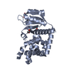
- Structure visualization
| Structure viewer | Molecule: MolmilJmol/JSmol |
|---|
- Downloads & links
-Download
| PDBx/mmCIF format | 4nxg.cif.gz | 106.1 KB | Display | PDBx/mmCIF format |
|---|---|---|---|---|
| PDB format | pdb4nxg.ent.gz | 80.1 KB | Display | PDB format |
| PDBx/mmJSON format | 4nxg.json.gz | Tree view | PDBx/mmJSON format | |
| Others |  Other downloads Other downloads |
-Validation report
| Arichive directory | https://data.pdbj.org/pub/pdb/validation_reports/nx/4nxgftp://data.pdbj.org/pub/pdb/validation_reports/nx/4nxg | HTTPS FTP |
|---|
-Related structure data
| Related structure data |  4nx2C 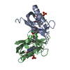 4nxbC 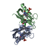 4nxeC 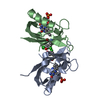 4nxfC 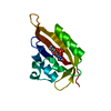 4eesS C: citing same article ( S: Starting model for refinement |
|---|---|
| Similar structure data |


-Links
 PDBj
PDBj


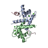
- Assembly
| Deposited unit | 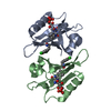
| ||||||||
|---|---|---|---|---|---|---|---|---|---|
| 1 |
| ||||||||
| Unit cell |
|
-Components
| #1: Protein | Mass: 13969.408 Da / Num. of mol.: 2 / Fragment: LOV DOMAIN, UNP Residues 388-496 / Mutation: S394T, S409G, I452T, F470L, M475V, I486Y Source method: isolated from a genetically manipulated source Source: (gene. exp.)  References: UniProt: P93025, non-specific serine/threonine protein kinase #2: Chemical |   Mass: 456.344 Da / Num. of mol.: 2 / Source method: obtained synthetically / Formula: C17H21N4O9P Mass: 456.344 Da / Num. of mol.: 2 / Source method: obtained synthetically / Formula: C17H21N4O9P#3: Water | ChemComp-HOH / |  Mass: 18.015 Da / Num. of mol.: 55 / Source method: isolated from a natural source / Formula: H2O Mass: 18.015 Da / Num. of mol.: 55 / Source method: isolated from a natural source / Formula: H2OSequence details | RESIDUES C426A IS MUTAGENESI | |
|---|
-Experimental details
-Experiment
| Experiment | Method: X-RAY DIFFRACTION / Number of used crystals: 1 |
|---|
- Sample preparation
| Crystal | Density Matthews: 1.95 Å3/Da / Density % sol: 36.85 % |
|---|---|
| Crystal grow | Temperature: 289 K / Method: vapor diffusion, sitting drop / pH: 9 Details: protein sample (20-30mg/ml) in 20mM Tris, pH 8.0, 50mM NaCl, equal volume of reservoir solution (0.1M BIS-TRIS propane pH 9.0, 30% w/v Polyethylene glycol 6000), VAPOR DIFFUSION, SITTING DROP, temperature 289.0K |
-Data collection
| Diffraction | Mean temperature: 200 K |
|---|---|
| Diffraction source | Source: SYNCHROTRON / Site: SSRF  / Beamline: BL17U / Wavelength: 0.979 Å / Beamline: BL17U / Wavelength: 0.979 Å |
| Detector | Type: ADSC QUANTUM 315 / Detector: CCD / Date: Jan 20, 2013 |
| Radiation | Protocol: SINGLE WAVELENGTH / Monochromatic (M) / Laue (L): M / Scattering type: x-ray |
| Radiation wavelength | Wavelength: 0.979 Å / Relative weight: 1 |
| Reflection | Resolution: 2.09→50 Å / Num. all: 12798 / Num. obs: 12632 / % possible obs: 98.7 % / Observed criterion σ(F): 1 / Observed criterion σ(I): 1 / Biso Wilson estimate: 27.12 Å2 |
| Reflection shell | Resolution: 2.09→2.13 Å / Redundancy: 3.7 % / Num. unique all: 586 / % possible all: 98 |
- Processing
| Software |
| ||||||||||||||||||||||||||||||||||||||||
|---|---|---|---|---|---|---|---|---|---|---|---|---|---|---|---|---|---|---|---|---|---|---|---|---|---|---|---|---|---|---|---|---|---|---|---|---|---|---|---|---|---|
| Refinement | Method to determine structure: MOLECULAR REPLACEMENT Starting model: 4EES Resolution: 2.09→36.375 Å / Occupancy max: 1 / Occupancy min: 0.48 / FOM work R set: 0.7945 / SU ML: 0.27 / σ(F): 1.35 / Phase error: 27.39 / Stereochemistry target values: ML
| ||||||||||||||||||||||||||||||||||||||||
| Solvent computation | Shrinkage radii: 0.9 Å / VDW probe radii: 1.11 Å / Solvent model: FLAT BULK SOLVENT MODEL | ||||||||||||||||||||||||||||||||||||||||
| Displacement parameters | Biso max: 65.07 Å2 / Biso mean: 26.0051 Å2 / Biso min: 10.29 Å2 | ||||||||||||||||||||||||||||||||||||||||
| Refinement step | Cycle: LAST / Resolution: 2.09→36.375 Å
| ||||||||||||||||||||||||||||||||||||||||
| Refine LS restraints |
| ||||||||||||||||||||||||||||||||||||||||
| LS refinement shell | Refine-ID: X-RAY DIFFRACTION / Total num. of bins used: 4
| ||||||||||||||||||||||||||||||||||||||||
| Refinement TLS params. | Method: refined / Origin x: -5.5138 Å / Origin y: -2.6549 Å / Origin z: 59.2752 Å
| ||||||||||||||||||||||||||||||||||||||||
| Refinement TLS group | Selection details: ALL |
