 Movie
Movie Controller


+ Open data

- Basic information
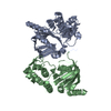
| Entry | Database: PDB / ID: 1quq | ||||||
|---|---|---|---|---|---|---|---|
| Title | COMPLEX OF REPLICATION PROTEIN A SUBUNITS RPA14 AND RPA32 | ||||||
 Components Components |
| ||||||
 Keywords Keywords | DNA BINDING PROTEIN / RPA / OB-FOLD / SSDNA-BINDING / DNA-BINDING PROTEIN | ||||||
| Function / homology |  Function and homology information Function and homology informationprotein localization to chromosome / DNA replication factor A complex / Removal of the Flap Intermediate / G-rich strand telomeric DNA binding / Mismatch repair (MMR) directed by MSH2:MSH3 (MutSbeta) / Mismatch repair (MMR) directed by MSH2:MSH6 (MutSalpha) / regulation of DNA damage checkpoint / Removal of the Flap Intermediate from the C-strand / regulation of double-strand break repair via homologous recombination / HDR through Single Strand Annealing (SSA) ...protein localization to chromosome / DNA replication factor A complex / Removal of the Flap Intermediate / G-rich strand telomeric DNA binding / Mismatch repair (MMR) directed by MSH2:MSH3 (MutSbeta) / Mismatch repair (MMR) directed by MSH2:MSH6 (MutSalpha) / regulation of DNA damage checkpoint / Removal of the Flap Intermediate from the C-strand / regulation of double-strand break repair via homologous recombination / HDR through Single Strand Annealing (SSA) / telomeric repeat DNA binding / Impaired BRCA2 binding to RAD51 / PCNA-Dependent Long Patch Base Excision Repair / Activation of the pre-replicative complex / Regulation of HSF1-mediated heat shock response / Presynaptic phase of homologous DNA pairing and strand exchange / HSF1 activation / Activation of ATR in response to replication stress / mismatch repair / mitotic G1 DNA damage checkpoint signaling / regulation of mitotic cell cycle / telomere maintenance / Translesion synthesis by REV1 / Translesion synthesis by POLK / Translesion synthesis by POLI / Gap-filling DNA repair synthesis and ligation in GG-NER / nucleotide-excision repair / Fanconi Anemia Pathway / Termination of translesion DNA synthesis / Translesion Synthesis by POLH / Recognition of DNA damage by PCNA-containing replication complex / base-excision repair / PML body / G2/M DNA damage checkpoint / double-strand break repair via homologous recombination / Meiotic recombination / HDR through Homologous Recombination (HRR) / Dual Incision in GG-NER / Formation of Incision Complex in GG-NER / Dual incision in TC-NER / Gap-filling DNA repair synthesis and ligation in TC-NER / regulation of cell population proliferation / single-stranded DNA binding / site of double-strand break / Processing of DNA double-strand break ends / protein phosphatase binding / Regulation of TP53 Activity through Phosphorylation / damaged DNA binding / DNA replication / chromosome, telomeric region / nuclear body / DNA repair / ubiquitin protein ligase binding / chromatin / enzyme binding / nucleoplasm / nucleus Similarity search - Function | ||||||
| Biological species |  Homo sapiens (human) Homo sapiens (human) | ||||||
| Method |  X-RAY DIFFRACTION / MAD / Resolution: 2.5 Å X-RAY DIFFRACTION / MAD / Resolution: 2.5 Å | ||||||
 Authors Authors | Bochkarev, A. / Bochkareva, E. / Frappier, L. / Edwards, A.M. | ||||||
 Citation Citation | Journal: EMBO J. / Year: 1999 Title: The crystal structure of the complex of replication protein A subunits RPA32 and RPA14 reveals a mechanism for single-stranded DNA binding. Authors: Bochkarev, A. / Bochkareva, E. / Frappier, L. / Edwards, A.M. #1: Journal: J.Biol.Chem. / Year: 1998Title: The Rpa32 Subunit of Human Replication Protein a Contains a Single-Stranded DNA-Binding Domain. Authors: Bochkareva, E. / Frappier, L. / Edwards, A.M. / Bochkarev, A. | ||||||
| History |
|
- Structure visualization
| Structure viewer | Molecule: MolmilJmol/JSmol |
|---|
- Downloads & links
-Download
| PDBx/mmCIF format | 1quq.cif.gz | 103 KB | Display | PDBx/mmCIF format |
|---|---|---|---|---|
| PDB format | pdb1quq.ent.gz | 80.7 KB | Display | PDB format |
| PDBx/mmJSON format | 1quq.json.gz | Tree view | PDBx/mmJSON format | |
| Others |  Other downloads Other downloads |
-Validation report
| Arichive directory | https://data.pdbj.org/pub/pdb/validation_reports/qu/1quqftp://data.pdbj.org/pub/pdb/validation_reports/qu/1quq | HTTPS FTP |
|---|
-Related structure data
| Similar structure data |
|---|


-Links
 PDBj
PDBj


- Assembly
| Deposited unit | 
| ||||||||
|---|---|---|---|---|---|---|---|---|---|
| 1 |
| ||||||||
| 2 | 
| ||||||||
| 3 | 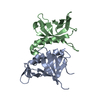
| ||||||||
| Unit cell |
|
-Components
| #1: Protein | Mass: 14432.592 Da / Num. of mol.: 2 / Fragment: CENTRAL DOMAIN, RESIDUES 43-171 Source method: isolated from a genetically manipulated source Source: (gene. exp.) Homo sapiens (human) / Cellular location: NUCLEUS / Plasmid: PET15B / Cell line (production host): BL21(DE3) / Production host:  #2: Protein | Mass: 13583.714 Da / Num. of mol.: 2 / Fragment: RPA14 Source method: isolated from a genetically manipulated source Source: (gene. exp.) Homo sapiens (human) / Cellular location: NUCLEUS / Plasmid: PET15B / Cell line (production host): BL21(DE3) / Production host: #3: Water | ChemComp-HOH / |  Mass: 18.015 Da / Num. of mol.: 117 / Source method: isolated from a natural source / Formula: H2O Mass: 18.015 Da / Num. of mol.: 117 / Source method: isolated from a natural source / Formula: H2O |
|---|
-Experimental details
-Experiment
| Experiment | Method: X-RAY DIFFRACTION / Number of used crystals: 1 |
|---|
- Sample preparation
| Crystal | Density Matthews: 2.68 Å3/Da / Density % sol: 54.1 % Description: MAD EXPERIMENT WAS COLLECTED AT CHESS BEAMLINE F2 IN APRIL 1998. DETECTOR - ADSC Q4. SOFTWARE - DENZO, SCALEPACK | ||||||||||||||||||||||||||||||||||||||||||||||||||||||
|---|---|---|---|---|---|---|---|---|---|---|---|---|---|---|---|---|---|---|---|---|---|---|---|---|---|---|---|---|---|---|---|---|---|---|---|---|---|---|---|---|---|---|---|---|---|---|---|---|---|---|---|---|---|---|---|
| Crystal grow | pH: 6.5 Details: 0.1 M MES, 0.75 M AMMONIUM SULPHATE, 20% PEG 8K, 10 MM DTT., pH 6.50 Temp details: 8 | ||||||||||||||||||||||||||||||||||||||||||||||||||||||
| Crystal grow | *PLUS pH: 7.5 / Method: vapor diffusion, hanging drop | ||||||||||||||||||||||||||||||||||||||||||||||||||||||
| Components of the solutions | *PLUS
|
-Data collection
| Diffraction | Mean temperature: 100 K |
|---|---|
| Diffraction source | Source: ROTATING ANODE / Type: RIGAKU RU300 / Wavelength: 1.5418 |
| Detector | Type: RIGAKU RAXIS IV++ / Detector: IMAGE PLATE / Date: Oct 1, 1998 / Details: OSMIC MULTILAYER |
| Radiation | Protocol: MAD / Monochromatic (M) / Laue (L): M / Scattering type: x-ray |
| Radiation wavelength | Wavelength: 1.5418 Å / Relative weight: 1 |
| Reflection | Resolution: 2.4→20 Å / Num. obs: 23882 / % possible obs: 98.6 % / Observed criterion σ(I): -3 / Redundancy: 5.2 % / Rmerge(I) obs: 0.044 / Net I/σ(I): 34.4 |
| Reflection shell | Resolution: 2.4→2.49 Å / Rsym value: 16.2 / % possible all: 96.9 |
| Reflection | *PLUS Highest resolution: 2.4 Å / Lowest resolution: 20 Å / Observed criterion σ(I): -3 / Redundancy: 5.2 % / Num. measured all: 123441 |
| Reflection shell | *PLUS % possible obs: 96.9 % / Rmerge(I) obs: 0.162 / Mean I/σ(I) obs: 7 |
- Processing
| Software |
| ||||||||||||||||||||||||||||||||||||||||||||||||||||||||||||||||||||||||||||||||
|---|---|---|---|---|---|---|---|---|---|---|---|---|---|---|---|---|---|---|---|---|---|---|---|---|---|---|---|---|---|---|---|---|---|---|---|---|---|---|---|---|---|---|---|---|---|---|---|---|---|---|---|---|---|---|---|---|---|---|---|---|---|---|---|---|---|---|---|---|---|---|---|---|---|---|---|---|---|---|---|---|---|
| Refinement | Method to determine structure: MAD / Resolution: 2.5→20 Å / Data cutoff high absF: 1000000 / Data cutoff low absF: 0.001 / Isotropic thermal model: RESTRAINED / Cross valid method: THROUGHOUT / σ(F): 2 Details: USED RESOLUTION DEPENDENT WEIGHTING AND BULK SOLVENT MODEL CORRECTION.
| ||||||||||||||||||||||||||||||||||||||||||||||||||||||||||||||||||||||||||||||||
| Displacement parameters | Biso mean: 22.1 Å2 | ||||||||||||||||||||||||||||||||||||||||||||||||||||||||||||||||||||||||||||||||
| Refinement step | Cycle: LAST / Resolution: 2.5→20 Å
| ||||||||||||||||||||||||||||||||||||||||||||||||||||||||||||||||||||||||||||||||
| Refine LS restraints |
| ||||||||||||||||||||||||||||||||||||||||||||||||||||||||||||||||||||||||||||||||
| LS refinement shell | Resolution: 2.5→2.61 Å / Total num. of bins used: 8
| ||||||||||||||||||||||||||||||||||||||||||||||||||||||||||||||||||||||||||||||||
| Xplor file |
| ||||||||||||||||||||||||||||||||||||||||||||||||||||||||||||||||||||||||||||||||
| Software | *PLUS Name: X-PLOR / Version: 3.843 / Classification: refinement | ||||||||||||||||||||||||||||||||||||||||||||||||||||||||||||||||||||||||||||||||
| Refinement | *PLUS Highest resolution: 2.5 Å / Lowest resolution: 20 Å / % reflection Rfree: 10 % | ||||||||||||||||||||||||||||||||||||||||||||||||||||||||||||||||||||||||||||||||
| Solvent computation | *PLUS | ||||||||||||||||||||||||||||||||||||||||||||||||||||||||||||||||||||||||||||||||
| Displacement parameters | *PLUS Biso mean: 22.1 Å2 | ||||||||||||||||||||||||||||||||||||||||||||||||||||||||||||||||||||||||||||||||
| Refine LS restraints | *PLUS
| ||||||||||||||||||||||||||||||||||||||||||||||||||||||||||||||||||||||||||||||||
| LS refinement shell | *PLUS Rfactor Rfree: 0.354 / % reflection Rfree: 8.7 % / Rfactor Rwork: 0.202 |
