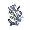
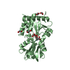
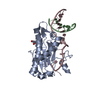
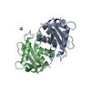
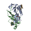
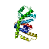
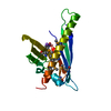
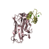
[English] 日本語
Yorodumi
- PDB-2y3n: Type II cohesin-dockerin domain from Bacteroides cellolosolvens -

+
Open data


ID or keywords:

Loading...

-
Basic information

Entry
Database: PDB / ID: 2y3n
TitleType II cohesin-dockerin domain from Bacteroides cellolosolvens
Components
  • CELLULOSOMAL FAMILY-48 PROCESSIVE GLYCOSIDE HYDROLASE
  • CELLULOSOMAL SCAFFOLDIN
KeywordsSTRUCTRUAL PROTEIN/HYDROLASE / STRUCTRUAL PROTEIN-HYDROLASE COMPLEX / CELLULOSOME
Function / homology
Function and homology information


cellulose 1,4-beta-cellobiosidase (non-reducing end) / cellulose 1,4-beta-cellobiosidase activity / cellulose binding / cellulase activity / cellulose catabolic process / polysaccharide catabolic process / hydrolase activity, hydrolyzing O-glycosyl compounds / extracellular region / metal ion binding
Similarity search - Function
Endoglucanase F, domain 2 / Endoglucanase F, domain 3 / Type 1 dockerin domain / Dockerin domain / Glycoside hydrolase, 48F / Glycosyl hydrolase family 48 / Immunoglobulin-like - #680 / Cellulosome anchoring protein, cohesin domain / Cohesin domain / Cellulose binding domain ...Endoglucanase F, domain 2 / Endoglucanase F, domain 3 / Type 1 dockerin domain / Dockerin domain / Glycoside hydrolase, 48F / Glycosyl hydrolase family 48 / Immunoglobulin-like - #680 / Cellulosome anchoring protein, cohesin domain / Cohesin domain / Cellulose binding domain / Cellulose binding domain / Carbohydrate-binding module 3 / Carbohydrate-binding module 3 superfamily / CBM3 (carbohydrate binding type-3) domain profile. / Dockerin domain / Dockerin domain profile. / Dockerin type I domain / Dockerin type I repeat / Dockerin domain superfamily / CBM2/CBM3, carbohydrate-binding domain superfamily / Six-hairpin glycosidase-like superfamily / Six-hairpin glycosidase superfamily / EF-Hand 1, calcium-binding site / EF-hand calcium-binding domain. / Immunoglobulin-like / Sandwich / Orthogonal Bundle / Mainly Beta / Mainly Alpha
Similarity search - Domain/homology
Cellulose 1,4-beta-cellobiosidase / Cellulosomal scaffoldin
Similarity search - Component
Biological speciesBACTEROIDES CELLULOSOLVENS (bacteria)
MethodX-RAY DIFFRACTION / SYNCHROTRON / MOLECULAR REPLACEMENT / Resolution: 1.9 Å
AuthorsViegas, A. / Carvalho, A.L. / Romao, M.J. / Prates, J.A.M. / Fontes, C.M.G.A.
CitationJournal: To be Published
Title: Type II Cohesin-Dockerin Domain from Bacteroides Cellolosolvens
Authors: Viegas, A. / Carvalho, A.L. / Prates, J.A.M. / Romao, M.J. / Fontes, C.M.G.A.
History
DepositionDec 22, 2010Deposition site: PDBE / Processing site: PDBE
Revision 1.0Jan 11, 2012Provider: repository / Type: Initial release
Revision 1.1Dec 20, 2023Group: Data collection / Database references ...Data collection / Database references / Derived calculations / Other / Refinement description
Category: chem_comp_atom / chem_comp_bond ...chem_comp_atom / chem_comp_bond / database_2 / pdbx_database_status / pdbx_initial_refinement_model / pdbx_struct_conn_angle / struct_conn / struct_site
Item: _database_2.pdbx_DOI / _database_2.pdbx_database_accession ..._database_2.pdbx_DOI / _database_2.pdbx_database_accession / _pdbx_database_status.status_code_sf / _pdbx_struct_conn_angle.ptnr1_auth_comp_id / _pdbx_struct_conn_angle.ptnr1_auth_seq_id / _pdbx_struct_conn_angle.ptnr1_label_asym_id / _pdbx_struct_conn_angle.ptnr1_label_atom_id / _pdbx_struct_conn_angle.ptnr1_label_comp_id / _pdbx_struct_conn_angle.ptnr1_label_seq_id / _pdbx_struct_conn_angle.ptnr2_auth_seq_id / _pdbx_struct_conn_angle.ptnr2_label_asym_id / _pdbx_struct_conn_angle.ptnr3_auth_comp_id / _pdbx_struct_conn_angle.ptnr3_auth_seq_id / _pdbx_struct_conn_angle.ptnr3_label_asym_id / _pdbx_struct_conn_angle.ptnr3_label_atom_id / _pdbx_struct_conn_angle.ptnr3_label_comp_id / _pdbx_struct_conn_angle.ptnr3_label_seq_id / _pdbx_struct_conn_angle.value / _struct_conn.pdbx_dist_value / _struct_conn.ptnr1_auth_comp_id / _struct_conn.ptnr1_auth_seq_id / _struct_conn.ptnr1_label_asym_id / _struct_conn.ptnr1_label_atom_id / _struct_conn.ptnr1_label_comp_id / _struct_conn.ptnr1_label_seq_id / _struct_conn.ptnr2_auth_comp_id / _struct_conn.ptnr2_auth_seq_id / _struct_conn.ptnr2_label_asym_id / _struct_conn.ptnr2_label_atom_id / _struct_conn.ptnr2_label_comp_id / _struct_conn.ptnr2_label_seq_id / _struct_site.pdbx_auth_asym_id / _struct_site.pdbx_auth_comp_id / _struct_site.pdbx_auth_seq_id

-
Structure visualization

Structure viewerMolecule:
MolmilJmol/JSmol

Downloads & links

-
Assembly

Deposited unit
A: CELLULOSOMAL SCAFFOLDIN
B: CELLULOSOMAL FAMILY-48 PROCESSIVE GLYCOSIDE HYDROLASE
C: CELLULOSOMAL SCAFFOLDIN
D: CELLULOSOMAL FAMILY-48 PROCESSIVE GLYCOSIDE HYDROLASE
hetero molecules


Theoretical massNumber of molelcules
Total (without water)55,3028
Polymers55,1424
Non-polymers1604
Water5,386299
1
C: CELLULOSOMAL SCAFFOLDIN
D: CELLULOSOMAL FAMILY-48 PROCESSIVE GLYCOSIDE HYDROLASE
hetero molecules


Theoretical massNumber of molelcules
Total (without water)27,6514
Polymers27,5712
Non-polymers802
Water362
TypeNameSymmetry operationNumber
identity operation1_555x,y,z1
Buried area1850 Å2
ΔGint-27 kcal/mol
Surface area10020 Å2
MethodPISA
2
A: CELLULOSOMAL SCAFFOLDIN
B: CELLULOSOMAL FAMILY-48 PROCESSIVE GLYCOSIDE HYDROLASE
hetero molecules


Theoretical massNumber of molelcules
Total (without water)27,6514
Polymers27,5712
Non-polymers802
Water362
TypeNameSymmetry operationNumber
identity operation1_555x,y,z1
Buried area1850 Å2
ΔGint-28.9 kcal/mol
Surface area10700 Å2
MethodPISA
Unit cell
Length a, b, c (Å)43.410, 116.120, 45.160
Angle α, β, γ (deg.)90.00, 112.45, 90.00
Int Tables number4
Space group name H-MP1211

-
Components

#1: Protein CELLULOSOMAL SCAFFOLDIN / TYPE II COHESIN-DOCKERIN


Mass: 20030.451 Da / Num. of mol.: 2 / Fragment: RESIDUES 2073-2242
Source method: isolated from a genetically manipulated source
Source: (gene. exp.) BACTEROIDES CELLULOSOLVENS (bacteria) / Production host: ESCHERICHIA COLI (E. coli) / Strain (production host): BL21 / References: UniProt: Q9FDJ9
#2: Protein CELLULOSOMAL FAMILY-48 PROCESSIVE GLYCOSIDE HYDROLASE / TYPE II COHESIN-DOCKERIN


Mass: 7540.627 Da / Num. of mol.: 2 / Fragment: RESIDUES 683-752
Source method: isolated from a genetically manipulated source
Source: (gene. exp.) BACTEROIDES CELLULOSOLVENS (bacteria) / Production host: ESCHERICHIA COLI (E. coli) / Strain (production host): BL21 / References: UniProt: Q6UB36
#3: Chemical
ChemComp-CA / CALCIUM ION


Mass: 40.078 Da / Num. of mol.: 4 / Source method: obtained synthetically / Formula: Ca
#4: Water ChemComp-HOH / water


Mass: 18.015 Da / Num. of mol.: 299 / Source method: isolated from a natural source / Formula: H2O

-
Experimental details

-
Experiment

ExperimentMethod: X-RAY DIFFRACTION / Number of used crystals: 1

-
Sample preparation

CrystalDensity Matthews: 1.91 Å3/Da / Density % sol: 35.67 % / Description: NONE

-
Data collection

DiffractionMean temperature: 287 K
Diffraction sourceSource: SYNCHROTRON / Site: ESRF / Beamline: ID23-1 / Wavelength: 0.9735
DetectorType: ADSC QUANTUM 315r / Detector: CCD
RadiationProtocol: SINGLE WAVELENGTH / Monochromatic (M) / Laue (L): M / Scattering type: x-ray
Radiation wavelengthWavelength: 0.9735 Å / Relative weight: 1
ReflectionResolution: 1.9→41.74 Å / Num. obs: 27197 / % possible obs: 83.7 % / Observed criterion σ(I): 2 / Redundancy: 2.4 % / Rmerge(I) obs: 0.09 / Net I/σ(I): 6.5
Reflection shellResolution: 1.9→41.74 Å / Redundancy: 2.2 % / Rmerge(I) obs: 0.33 / Mean I/σ(I) obs: 3.2 / % possible all: 83.7

-
Processing

Software
NameVersionClassification
REFMAC5.5.0110refinement
MOSFLMdata reduction
SCALAdata scaling
PHASERphasing
RefinementMethod to determine structure: MOLECULAR REPLACEMENT
Starting model: PDB ENTRY 1TYJ
Resolution: 1.9→41.74 Å / Cor.coef. Fo:Fc: 0.959 / Cor.coef. Fo:Fc free: 0.927 / SU B: 7.713 / SU ML: 0.106 / Cross valid method: THROUGHOUT / ESU R: 0.198 / ESU R Free: 0.176 / Stereochemistry target values: MAXIMUM LIKELIHOOD / Details: HYDROGENS HAVE BEEN ADDED IN THE RIDING POSITIONS.
RfactorNum. reflection% reflectionSelection details
Rfree0.22491 1364 5 %RANDOM
Rwork0.16329 ---
obs0.16641 25833 83.7 %-
Solvent computationIon probe radii: 0.8 Å / Shrinkage radii: 0.8 Å / VDW probe radii: 1.4 Å / Solvent model: MASK
Displacement parametersBiso mean: 27.086 Å2
Baniso -1Baniso -2Baniso -3
1--0.03 Å20 Å20.28 Å2
2--0 Å20 Å2
3---0.25 Å2
Refinement stepCycle: LAST / Resolution: 1.9→41.74 Å
ProteinNucleic acidLigandSolventTotal
Num. atoms3462 0 4 299 3765
Refine LS restraints
Refine-IDTypeDev idealDev ideal targetNumber
X-RAY DIFFRACTIONr_bond_refined_d0.0220.0223520
X-RAY DIFFRACTIONr_bond_other_d
X-RAY DIFFRACTIONr_angle_refined_deg1.6961.9554758
X-RAY DIFFRACTIONr_angle_other_deg
X-RAY DIFFRACTIONr_dihedral_angle_1_deg6.8665447
X-RAY DIFFRACTIONr_dihedral_angle_2_deg35.01526.026151
X-RAY DIFFRACTIONr_dihedral_angle_3_deg15.01215602
X-RAY DIFFRACTIONr_dihedral_angle_4_deg13.542156
X-RAY DIFFRACTIONr_chiral_restr0.1240.2540
X-RAY DIFFRACTIONr_gen_planes_refined0.010.0212622
X-RAY DIFFRACTIONr_gen_planes_other
X-RAY DIFFRACTIONr_nbd_refined
X-RAY DIFFRACTIONr_nbd_other
X-RAY DIFFRACTIONr_nbtor_refined
X-RAY DIFFRACTIONr_nbtor_other
X-RAY DIFFRACTIONr_xyhbond_nbd_refined
X-RAY DIFFRACTIONr_xyhbond_nbd_other
X-RAY DIFFRACTIONr_metal_ion_refined
X-RAY DIFFRACTIONr_metal_ion_other
X-RAY DIFFRACTIONr_symmetry_vdw_refined
X-RAY DIFFRACTIONr_symmetry_vdw_other
X-RAY DIFFRACTIONr_symmetry_hbond_refined
X-RAY DIFFRACTIONr_symmetry_hbond_other
X-RAY DIFFRACTIONr_symmetry_metal_ion_refined
X-RAY DIFFRACTIONr_symmetry_metal_ion_other
X-RAY DIFFRACTIONr_mcbond_it1.41.52231
X-RAY DIFFRACTIONr_mcbond_other
X-RAY DIFFRACTIONr_mcangle_it2.29223577
X-RAY DIFFRACTIONr_mcangle_other
X-RAY DIFFRACTIONr_scbond_it3.57531289
X-RAY DIFFRACTIONr_scbond_other
X-RAY DIFFRACTIONr_scangle_it5.4114.51181
X-RAY DIFFRACTIONr_scangle_other
X-RAY DIFFRACTIONr_long_range_B_refined
X-RAY DIFFRACTIONr_long_range_B_other
X-RAY DIFFRACTIONr_rigid_bond_restr
X-RAY DIFFRACTIONr_sphericity_free
X-RAY DIFFRACTIONr_sphericity_bonded
LS refinement shellResolution: 1.9→1.949 Å / Total num. of bins used: 20
RfactorNum. reflection% reflection
Rfree0.218 67 -
Rwork0.165 1272 -
obs--56.83 %
Refinement TLS params.

Method: refined / Refine-ID: X-RAY DIFFRACTION

IDL112)L122)L132)L222)L232)L332)S11 (Å °)S12 (Å °)S13 (Å °)S21 (Å °)S22 (Å °)S23 (Å °)S31 (Å °)S32 (Å °)S33 (Å °)T112)T122)T132)T222)T232)T332)Origin x (Å)Origin y (Å)Origin z (Å)
10.528-0.10870.74460.3841-0.4191.18940.03140.05010.0077-0.0674-0.0192-0.0260.03160.0428-0.01220.15510.0039-0.02170.12410.00250.0516-13.24426.195-20.378
20.4548-0.15610.3260.3254-0.34990.8020.0215-0.006-0.0436-0.03860.00060.01520.0512-0.064-0.02220.15350.0007-0.02560.10450.00370.0654-14.55821.324-13.606
30.9992-0.26730.57060.6041-0.34050.67910.02470.1361-0.0048-0.0452-0.0032-0.0647-0.12560.1996-0.02150.1445-0.0083-0.02640.106-0.00450.0728-9.68531.249-18.094
48.8775-2.2091-5.97017.74992.8943.3889-0.0557-0.22940.01480.06130.1543-0.05990.03030.1446-0.09860.17360.0522-0.04280.14650.01290.0224-23.96126.30810.858
51.6475-1.9998-0.12672.08240.14872.3257-0.0789-0.11810.00780.12760.0872-0.0015-0.15150.042-0.00820.1816-0.0481-0.00790.1435-0.0210.0525-13.9621.6353.752
62.9778-2.3736-4.90033.03046.28328.0349-0.2795-1.1688-0.28140.44240.171-0.06870.1130.50790.10850.45110.0643-0.07490.4410.11350.0067-15.18111.10611.76
72.4167-3.85820.43569.1412-1.49221.174-0.0437-0.0674-0.04390.03180.22710.3757-0.1357-0.2731-0.18340.11120.02260.0110.10980.0340.0823-21.69718.4582.171
81.04960.30290.05910.5996-0.11480.2878-0.0048-0.02350.05620.00190.00460.0169-0.0022-0.00490.00020.1112-0.0049-0.01990.0979-0.01540.1097-3.017-4.8192.659
91.73390.5833-0.90290.37110.0050.8105-0.01110.0826-0.02720.02720.047-0.01990.0475-0.0572-0.0360.0971-0.0261-0.01370.11280.00750.1226-8.915-18.9243.29
100.64510.87560.07551.4456-0.20470.40530.02190.0204-0.09920.0050.0185-0.20060.08130.0768-0.04040.0845-0.0022-0.01730.1156-0.00280.12955.007-13.2413.088
116.2932-2.0481-1.06859.5114-4.19793.71830.0403-0.23080.27211.3975-0.0970.56070.57480.60520.05670.50640.07630.16010.1838-0.02870.03559.691-10.383-23.761
121.14842.64941.06266.17143.97942.118-0.13720.1561-0.0451-0.64440.4819-0.4764-0.38350.2385-0.34470.1767-0.05480.09910.1726-0.05590.160511.583-4.353-11.344
1320.126717.65857.668814.52715.30932.2046-0.45360.26541.0027-0.94920.26760.0794-0.1797-0.16130.18590.45350.09630.25930.1570.07530.30687.9824.48-14.352
146.5433-16.2523-17.393924.4721-0.554223.88260.58860.39941.6153-1.241.2591-2.72620.1531-2.7571-1.84770.0583-0.0880.08080.2773-0.00460.38862.147-9.998-16.006
Refinement TLS group
IDRefine-IDRefine TLS-IDAuth asym-IDAuth seq-ID
1X-RAY DIFFRACTION1A3 - 48
2X-RAY DIFFRACTION2A49 - 152
3X-RAY DIFFRACTION3A153 - 175
4X-RAY DIFFRACTION4B1 - 7
5X-RAY DIFFRACTION5B8 - 27
6X-RAY DIFFRACTION6B28 - 47
7X-RAY DIFFRACTION7B48 - 66
8X-RAY DIFFRACTION8C3 - 123
9X-RAY DIFFRACTION9C124 - 138
10X-RAY DIFFRACTION10C139 - 173
11X-RAY DIFFRACTION11D1 - 6
12X-RAY DIFFRACTION12D7 - 28
13X-RAY DIFFRACTION13D29 - 59
14X-RAY DIFFRACTION14D60 - 64

+
About Yorodumi

-
News

-
Feb 9, 2022. New format data for meta-information of EMDB entries

New format data for meta-information of EMDB entries

  • Version 3 of the EMDB header file is now the official format.
  • The previous official version 1.9 will be removed from the archive.

Related info.:EMDB header

External links:wwPDB to switch to version 3 of the EMDB data model

-
Aug 12, 2020. Covid-19 info

Covid-19 info

URL: https://pdbjlvh1.pdbj.org/emnavi/covid19.php

New page: Covid-19 featured information page in EM Navigator.

Related info.:Covid-19 info / Mar 5, 2020. Novel coronavirus structure data

+
Mar 5, 2020. Novel coronavirus structure data

Novel coronavirus structure data

Related info.:Yorodumi Speices / Aug 12, 2020. Covid-19 info

External links:COVID-19 featured content - PDBj / Molecule of the Month (242):Coronavirus Proteases

+
Jan 31, 2019. EMDB accession codes are about to change! (news from PDBe EMDB page)

EMDB accession codes are about to change! (news from PDBe EMDB page)

  • The allocation of 4 digits for EMDB accession codes will soon come to an end. Whilst these codes will remain in use, new EMDB accession codes will include an additional digit and will expand incrementally as the available range of codes is exhausted. The current 4-digit format prefixed with “EMD-” (i.e. EMD-XXXX) will advance to a 5-digit format (i.e. EMD-XXXXX), and so on. It is currently estimated that the 4-digit codes will be depleted around Spring 2019, at which point the 5-digit format will come into force.
  • The EM Navigator/Yorodumi systems omit the EMD- prefix.

Related info.:Q: What is EMD? / ID/Accession-code notation in Yorodumi/EM Navigator

External links:EMDB Accession Codes are Changing Soon! / Contact to PDBj

+
Jul 12, 2017. Major update of PDB

Major update of PDB

  • wwPDB released updated PDB data conforming to the new PDBx/mmCIF dictionary.
  • This is a major update changing the version number from 4 to 5, and with Remediation, in which all the entries are updated.
  • In this update, many items about electron microscopy experimental information are reorganized (e.g. em_software).
  • Now, EM Navigator and Yorodumi are based on the updated data.

External links:wwPDB Remediation / Enriched Model Files Conforming to OneDep Data Standards Now Available in the PDB FTP Archive

-
Yorodumi

Thousand views of thousand structures

  • Yorodumi is a browser for structure data from EMDB, PDB, SASBDB, etc.
  • This page is also the successor to EM Navigator detail page, and also detail information page/front-end page for Omokage search.
  • The word "yorodu" (or yorozu) is an old Japanese word meaning "ten thousand". "mi" (miru) is to see.

Related info.:EMDB / PDB / SASBDB / Comparison of 3 databanks / Yorodumi Search / Aug 31, 2016. New EM Navigator & Yorodumi / Yorodumi Papers / Jmol/JSmol / Function and homology information / Changes in new EM Navigator and Yorodumi

Read more