 Movie
Movie Controller

[English] 日本語
 Yorodumi
- PDB-3ijf: Crystal structure of cytidine deaminase from Mycobacterium tuberc... -
+ Open data

- Basic information
| Entry | Database: PDB / ID: 3ijf | ||||||
|---|---|---|---|---|---|---|---|
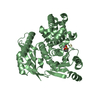
| Title | Crystal structure of cytidine deaminase from Mycobacterium tuberculosis | ||||||
 Components Components | Cytidine deaminase | ||||||
 Keywords Keywords | HYDROLASE / Mycobacterium tuberculosis / cytidine deaminase / drug target | ||||||
| Function / homology |  Function and homology information Function and homology informationuridine biosynthetic process / pyrimidine nucleobase salvage / cytidine deaminase / cytidine deaminase activity / zinc ion binding / metal ion binding / plasma membrane / cytosol Similarity search - Function | ||||||
| Biological species |   Mycobacterium tuberculosis (bacteria) Mycobacterium tuberculosis (bacteria) | ||||||
| Method |  X-RAY DIFFRACTION / SYNCHROTRON / MOLECULAR REPLACEMENT / Resolution: 1.99 Å X-RAY DIFFRACTION / SYNCHROTRON / MOLECULAR REPLACEMENT / Resolution: 1.99 Å | ||||||
 Authors Authors | De Azevedo Jr., W.F. / Basso, L.A. / Santos, D.S. | ||||||
 Citation Citation | Journal: J.Struct.Biol. / Year: 2010 Title: Structural and functional analyses of Mycobacterium tuberculosis Rv3315c-encoded metal-dependent homotetrameric cytidine deaminase. Authors: Sanchez-Quitian, Z.A. / Schneider, C.Z. / Ducati, R.G. / de Azevedo, W.F. / Bloch, C. / Basso, L.A. / Santos, D.S. | ||||||
| History |
|
- Structure visualization
| Structure viewer | Molecule: MolmilJmol/JSmol |
|---|
- Downloads & links
-Download
| PDBx/mmCIF format | 3ijf.cif.gz | 38.1 KB | Display | PDBx/mmCIF format |
|---|---|---|---|---|
| PDB format | pdb3ijf.ent.gz | 25.2 KB | Display | PDB format |
| PDBx/mmJSON format | 3ijf.json.gz | Tree view | PDBx/mmJSON format | |
| Others |  Other downloads Other downloads |
-Validation report
| Arichive directory | https://data.pdbj.org/pub/pdb/validation_reports/ij/3ijfftp://data.pdbj.org/pub/pdb/validation_reports/ij/3ijf | HTTPS FTP |
|---|
-Related structure data
| Related structure data |  2fr5S S: Starting model for refinement |
|---|---|
| Similar structure data |

-Links
 PDBj
PDBj- Assembly
| Deposited unit | 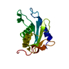
| ||||||||
|---|---|---|---|---|---|---|---|---|---|
| 1 | 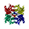
| ||||||||
| 2 | 
| ||||||||
| 3 | 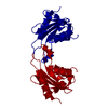
| ||||||||
| Unit cell |
|
-Components
| #1: Protein | Mass: 14088.142 Da / Num. of mol.: 1 Source method: isolated from a genetically manipulated source Source: (gene. exp.) Mycobacterium tuberculosis (bacteria) / Strain: H37Rv / Gene: cdd, MT3416, Rv3315c / Plasmid: pET-23a(+) / Production host: References: UniProt: O53367, UniProt: P9WPH3*PLUS, cytidine deaminase |
|---|---|
| #2: Chemical | ChemComp-ZN /   Mass: 65.409 Da / Num. of mol.: 1 / Source method: obtained synthetically / Formula: Zn Mass: 65.409 Da / Num. of mol.: 1 / Source method: obtained synthetically / Formula: Zn |
| #3: Water | ChemComp-HOH /  Mass: 18.015 Da / Num. of mol.: 89 / Source method: isolated from a natural source / Formula: H2O Mass: 18.015 Da / Num. of mol.: 89 / Source method: isolated from a natural source / Formula: H2O |
-Experimental details
-Experiment
| Experiment | Method: X-RAY DIFFRACTION / Number of used crystals: 1 |
|---|
- Sample preparation
| Crystal | Density Matthews: 2.35 Å3/Da / Density % sol: 47.59 % |
|---|---|
| Crystal grow | Temperature: 293 K / Method: vapor diffusion, hanging drop / pH: 7.5 Details: 100 mM HEPES and 4.3 M NaCl, pH 7.5, VAPOR DIFFUSION, HANGING DROP, temperature 293K |
-Data collection
| Diffraction | Mean temperature: 100 K |
|---|---|
| Diffraction source | Source: SYNCHROTRON / Site: LNLS  / Beamline: D03B-MX1 / Wavelength: 1.437 Å / Beamline: D03B-MX1 / Wavelength: 1.437 Å |
| Detector | Type: MAC Science DIP-3000 / Detector: IMAGE PLATE / Date: Feb 7, 2009 |
| Radiation | Monochromator: GRAPHITE / Protocol: SINGLE WAVELENGTH / Monochromatic (M) / Laue (L): M / Scattering type: x-ray |
| Radiation wavelength | Wavelength: 1.437 Å / Relative weight: 1 |
| Reflection | Resolution: 1.99→22.27 Å / Num. all: 20845 / Num. obs: 9374 / % possible obs: 97 % / Observed criterion σ(F): 2 / Observed criterion σ(I): 2 / Redundancy: 2.3 % / Rmerge(I) obs: 0.086 / Rsym value: 0.086 / Net I/σ(I): 10 |
- Processing
| Software |
| ||||||||||||||||||||||||||||||||||||||||||||||||||||||||||||||||||||||||||||||||||||||||||||||||||||||||||||||||||||||||||||||||||||||||||||||||||||||||||||||||||||||||||
|---|---|---|---|---|---|---|---|---|---|---|---|---|---|---|---|---|---|---|---|---|---|---|---|---|---|---|---|---|---|---|---|---|---|---|---|---|---|---|---|---|---|---|---|---|---|---|---|---|---|---|---|---|---|---|---|---|---|---|---|---|---|---|---|---|---|---|---|---|---|---|---|---|---|---|---|---|---|---|---|---|---|---|---|---|---|---|---|---|---|---|---|---|---|---|---|---|---|---|---|---|---|---|---|---|---|---|---|---|---|---|---|---|---|---|---|---|---|---|---|---|---|---|---|---|---|---|---|---|---|---|---|---|---|---|---|---|---|---|---|---|---|---|---|---|---|---|---|---|---|---|---|---|---|---|---|---|---|---|---|---|---|---|---|---|---|---|---|---|---|---|---|
| Refinement | Method to determine structure: MOLECULAR REPLACEMENT Starting model: 2fr5 Resolution: 1.99→18.24 Å / Cor.coef. Fo:Fc: 0.937 / Cor.coef. Fo:Fc free: 0.909 / SU B: 4.18 / SU ML: 0.11 / Cross valid method: THROUGHOUT / ESU R: 0.17 / ESU R Free: 0.16 / Stereochemistry target values: MAXIMUM LIKELIHOOD / Details: HYDROGENS HAVE BEEN ADDED IN THE RIDING POSITIONS
| ||||||||||||||||||||||||||||||||||||||||||||||||||||||||||||||||||||||||||||||||||||||||||||||||||||||||||||||||||||||||||||||||||||||||||||||||||||||||||||||||||||||||||
| Solvent computation | Ion probe radii: 0.8 Å / Shrinkage radii: 0.8 Å / VDW probe radii: 1.2 Å / Solvent model: MASK | ||||||||||||||||||||||||||||||||||||||||||||||||||||||||||||||||||||||||||||||||||||||||||||||||||||||||||||||||||||||||||||||||||||||||||||||||||||||||||||||||||||||||||
| Displacement parameters | Biso mean: 17.537 Å2
| ||||||||||||||||||||||||||||||||||||||||||||||||||||||||||||||||||||||||||||||||||||||||||||||||||||||||||||||||||||||||||||||||||||||||||||||||||||||||||||||||||||||||||
| Refinement step | Cycle: LAST / Resolution: 1.99→18.24 Å
| ||||||||||||||||||||||||||||||||||||||||||||||||||||||||||||||||||||||||||||||||||||||||||||||||||||||||||||||||||||||||||||||||||||||||||||||||||||||||||||||||||||||||||
| Refine LS restraints |
| ||||||||||||||||||||||||||||||||||||||||||||||||||||||||||||||||||||||||||||||||||||||||||||||||||||||||||||||||||||||||||||||||||||||||||||||||||||||||||||||||||||||||||
| LS refinement shell | Resolution: 1.994→2.046 Å / Total num. of bins used: 20
|
