 Movie
Movie Controller

[English] 日本語
 Yorodumi
- PDB-5ex5: Crystal structure of human GRP78 (70kDa heat shock protein 5 / BI... -
+ Open data

- Basic information
| Entry | Database: PDB / ID: 5ex5 | ||||||
|---|---|---|---|---|---|---|---|
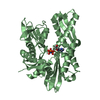
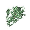
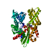
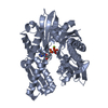
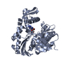
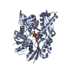
| Title | Crystal structure of human GRP78 (70kDa heat shock protein 5 / BIP) ATPase domain in complex with 7-deaza-ADP and inorganic phosphate | ||||||
 Components Components | 78 kDa glucose-regulated protein | ||||||
 Keywords Keywords | CHAPERONE / ATPase domain / nucleotide-binding / endoplasmic reticulum | ||||||
| Function / homology |  Function and homology information Function and homology informationregulation of ATF6-mediated unfolded protein response / regulation of PERK-mediated unfolded protein response / regulation of protein folding in endoplasmic reticulum / cerebellum structural organization / ATF6 (ATF6-alpha) activates chaperones / ATF6B (ATF6-beta) activates chaperones / maintenance of protein localization in endoplasmic reticulum / IRE1alpha activates chaperones / ATF6 (ATF6-alpha) activates chaperone genes / endoplasmic reticulum chaperone complex ...regulation of ATF6-mediated unfolded protein response / regulation of PERK-mediated unfolded protein response / regulation of protein folding in endoplasmic reticulum / cerebellum structural organization / ATF6 (ATF6-alpha) activates chaperones / ATF6B (ATF6-beta) activates chaperones / maintenance of protein localization in endoplasmic reticulum / IRE1alpha activates chaperones / ATF6 (ATF6-alpha) activates chaperone genes / endoplasmic reticulum chaperone complex / negative regulation of IRE1-mediated unfolded protein response / regulation of IRE1-mediated unfolded protein response / PERK regulates gene expression / protein folding in endoplasmic reticulum / cerebellar Purkinje cell layer development / misfolded protein binding / post-translational protein targeting to membrane, translocation / Modulation of host responses by IFN-stimulated genes / ER overload response / IRE1-mediated unfolded protein response / negative regulation of PERK-mediated unfolded protein response / endoplasmic reticulum-Golgi intermediate compartment / non-chaperonin molecular chaperone ATPase / intracellular membrane-bounded organelle / Regulation of HSF1-mediated heat shock response / protein serine/threonine kinase inhibitor activity / negative regulation of protein-containing complex assembly / cellular response to glucose starvation / cellular response to interleukin-4 / endoplasmic reticulum unfolded protein response / heat shock protein binding / ERAD pathway / protein folding chaperone / substantia nigra development / response to endoplasmic reticulum stress / positive regulation of protein ubiquitination / Antigen Presentation: Folding, assembly and peptide loading of class I MHC / ATP-dependent protein folding chaperone / protein sequestering activity / negative regulation of transforming growth factor beta receptor signaling pathway / : / melanosome / Platelet degranulation / protein refolding / protein-folding chaperone binding / ribosome binding / midbody / positive regulation of cell migration / cadherin binding / endoplasmic reticulum lumen / protein domain specific binding / focal adhesion / calcium ion binding / ubiquitin protein ligase binding / negative regulation of apoptotic process / endoplasmic reticulum membrane / enzyme binding / cell surface / endoplasmic reticulum / positive regulation of transcription by RNA polymerase II / ATP hydrolysis activity / protein-containing complex / mitochondrion / extracellular exosome / ATP binding / membrane / nucleus / plasma membrane / cytosol / cytoplasm Similarity search - Function | ||||||
| Biological species |  Homo sapiens (human) Homo sapiens (human) | ||||||
| Method |  X-RAY DIFFRACTION / SYNCHROTRON / MOLECULAR REPLACEMENT / Resolution: 1.9 Å X-RAY DIFFRACTION / SYNCHROTRON / MOLECULAR REPLACEMENT / Resolution: 1.9 Å | ||||||
 Authors Authors | Hughes, S.J. / Antoshchenko, T. / Song, J.H. / Pizarro, J. / Park, H.W. | ||||||
 Citation Citation | Journal: Plos One / Year: 2016 Title: Probing the ATP Site of GRP78 with Nucleotide Triphosphate Analogs. Authors: Hughes, S.J. / Antoshchenko, T. / Chen, Y. / Lu, H. / Pizarro, J.C. / Park, H.W. | ||||||
| History |
|
- Structure visualization
| Structure viewer | Molecule: MolmilJmol/JSmol |
|---|
- Downloads & links
-Download
| PDBx/mmCIF format | 5ex5.cif.gz | 319.2 KB | Display | PDBx/mmCIF format |
|---|---|---|---|---|
| PDB format | pdb5ex5.ent.gz | 257 KB | Display | PDB format |
| PDBx/mmJSON format | 5ex5.json.gz | Tree view | PDBx/mmJSON format | |
| Others |  Other downloads Other downloads |
-Validation report
| Arichive directory | https://data.pdbj.org/pub/pdb/validation_reports/ex/5ex5ftp://data.pdbj.org/pub/pdb/validation_reports/ex/5ex5 | HTTPS FTP |
|---|
-Related structure data
| Related structure data | 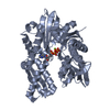 5evzC 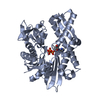 5exwSC 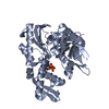 5ey4C 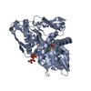 5f0xC 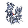 5f1xC  5f2rC C: citing same article ( S: Starting model for refinement |
|---|---|
| Similar structure data |


-Links
 PDBj
PDBj


- Assembly
| Deposited unit | 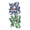
| ||||||||
|---|---|---|---|---|---|---|---|---|---|
| 1 | 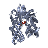
| ||||||||
| 2 | 
| ||||||||
| Unit cell |
|
-Components
| #1: Protein | Mass: 44356.090 Da / Num. of mol.: 2 / Fragment: ATPase domain (UNP residues 26-407) Source method: isolated from a genetically manipulated source Source: (gene. exp.) Homo sapiens (human) / Gene: HSPA5, GRP78 / Production host:  #2: Chemical |   Mass: 24.305 Da / Num. of mol.: 2 / Source method: obtained synthetically / Formula: Mg Mass: 24.305 Da / Num. of mol.: 2 / Source method: obtained synthetically / Formula: Mg#3: Chemical |   Mass: 94.971 Da / Num. of mol.: 2 / Source method: obtained synthetically / Formula: PO4 Mass: 94.971 Da / Num. of mol.: 2 / Source method: obtained synthetically / Formula: PO4#4: Chemical |   Mass: 426.213 Da / Num. of mol.: 2 / Source method: obtained synthetically / Formula: C11H16N4O10P2 Mass: 426.213 Da / Num. of mol.: 2 / Source method: obtained synthetically / Formula: C11H16N4O10P2#5: Water | ChemComp-HOH / |  Mass: 18.015 Da / Num. of mol.: 500 / Source method: isolated from a natural source / Formula: H2O Mass: 18.015 Da / Num. of mol.: 500 / Source method: isolated from a natural source / Formula: H2O |
|---|
-Experimental details
-Experiment
| Experiment | Method: X-RAY DIFFRACTION / Number of used crystals: 1 |
|---|
- Sample preparation
| Crystal | Density Matthews: 2.04 Å3/Da / Density % sol: 39.79 % |
|---|---|
| Crystal grow | Temperature: 289 K / Method: vapor diffusion, hanging drop / pH: 8.5 Details: 24-26% PEG3350, 0.1 M Tris-HCl, 0.2 M sodium chloride |
-Data collection
| Diffraction | Mean temperature: 100 K | ||||||||||||||||||||||||||||||
|---|---|---|---|---|---|---|---|---|---|---|---|---|---|---|---|---|---|---|---|---|---|---|---|---|---|---|---|---|---|---|---|
| Diffraction source | Source: SYNCHROTRON / Site: CLSI  / Beamline: 08ID-1 / Wavelength: 0.97949 Å / Beamline: 08ID-1 / Wavelength: 0.97949 Å | ||||||||||||||||||||||||||||||
| Detector | Type: RAYONIX MX-300 / Detector: CCD / Date: Oct 1, 2015 | ||||||||||||||||||||||||||||||
| Radiation | Monochromator: Double crystal Si(111) / Protocol: SINGLE WAVELENGTH / Monochromatic (M) / Laue (L): M / Scattering type: x-ray | ||||||||||||||||||||||||||||||
| Radiation wavelength | Wavelength: 0.97949 Å / Relative weight: 1 | ||||||||||||||||||||||||||||||
| Reflection | Resolution: 1.9→40 Å / Num. all: 55831 / Num. obs: 55831 / % possible obs: 99.1 % / Redundancy: 4.2 % / CC1/2: 0.993 / Rmerge(I) obs: 0.117 / Rpim(I) all: 0.064 / Rrim(I) all: 0.134 / Rsym value: 0.117 / Net I/σ(I): 9.3 / Num. measured all: 234113 | ||||||||||||||||||||||||||||||
| Reflection shell | Diffraction-ID: 1 / Rejects: _
|
- Processing
| Software |
| |||||||||||||||||||||||||||||||||||||||||||||||||||||||||||||||||||||||||||||||||||||||||||||||||||||||||||||||||||||||||||||||||||||||||||||||||||||||||||||||||||||||||||||||||||||||||||||||||||||||||||||||||||||||||||||||||
|---|---|---|---|---|---|---|---|---|---|---|---|---|---|---|---|---|---|---|---|---|---|---|---|---|---|---|---|---|---|---|---|---|---|---|---|---|---|---|---|---|---|---|---|---|---|---|---|---|---|---|---|---|---|---|---|---|---|---|---|---|---|---|---|---|---|---|---|---|---|---|---|---|---|---|---|---|---|---|---|---|---|---|---|---|---|---|---|---|---|---|---|---|---|---|---|---|---|---|---|---|---|---|---|---|---|---|---|---|---|---|---|---|---|---|---|---|---|---|---|---|---|---|---|---|---|---|---|---|---|---|---|---|---|---|---|---|---|---|---|---|---|---|---|---|---|---|---|---|---|---|---|---|---|---|---|---|---|---|---|---|---|---|---|---|---|---|---|---|---|---|---|---|---|---|---|---|---|---|---|---|---|---|---|---|---|---|---|---|---|---|---|---|---|---|---|---|---|---|---|---|---|---|---|---|---|---|---|---|---|---|---|---|---|---|---|---|---|---|---|---|---|---|---|---|---|---|
| Refinement | Method to determine structure: MOLECULAR REPLACEMENT Starting model: PDB entry 5EXW Resolution: 1.9→37.87 Å / Cor.coef. Fo:Fc: 0.955 / Cor.coef. Fo:Fc free: 0.936 / WRfactor Rfree: 0.2119 / WRfactor Rwork: 0.1685 / FOM work R set: 0.884 / SU B: 6.001 / SU ML: 0.099 / SU R Cruickshank DPI: 0.1628 / SU Rfree: 0.1443 / Cross valid method: THROUGHOUT / σ(F): 0 / ESU R: 0.163 / ESU R Free: 0.144 / Stereochemistry target values: MAXIMUM LIKELIHOOD / Details: HYDROGENS HAVE BEEN ADDED IN THE RIDING POSITIONS
| |||||||||||||||||||||||||||||||||||||||||||||||||||||||||||||||||||||||||||||||||||||||||||||||||||||||||||||||||||||||||||||||||||||||||||||||||||||||||||||||||||||||||||||||||||||||||||||||||||||||||||||||||||||||||||||||||
| Solvent computation | Ion probe radii: 0.7 Å / Shrinkage radii: 0.7 Å / VDW probe radii: 1.2 Å / Solvent model: MASK | |||||||||||||||||||||||||||||||||||||||||||||||||||||||||||||||||||||||||||||||||||||||||||||||||||||||||||||||||||||||||||||||||||||||||||||||||||||||||||||||||||||||||||||||||||||||||||||||||||||||||||||||||||||||||||||||||
| Displacement parameters | Biso max: 87.38 Å2 / Biso mean: 25.438 Å2 / Biso min: 4.67 Å2
| |||||||||||||||||||||||||||||||||||||||||||||||||||||||||||||||||||||||||||||||||||||||||||||||||||||||||||||||||||||||||||||||||||||||||||||||||||||||||||||||||||||||||||||||||||||||||||||||||||||||||||||||||||||||||||||||||
| Refinement step | Cycle: final / Resolution: 1.9→37.87 Å
| |||||||||||||||||||||||||||||||||||||||||||||||||||||||||||||||||||||||||||||||||||||||||||||||||||||||||||||||||||||||||||||||||||||||||||||||||||||||||||||||||||||||||||||||||||||||||||||||||||||||||||||||||||||||||||||||||
| Refine LS restraints |
| |||||||||||||||||||||||||||||||||||||||||||||||||||||||||||||||||||||||||||||||||||||||||||||||||||||||||||||||||||||||||||||||||||||||||||||||||||||||||||||||||||||||||||||||||||||||||||||||||||||||||||||||||||||||||||||||||
| LS refinement shell | Resolution: 1.9→1.949 Å / Total num. of bins used: 20
| |||||||||||||||||||||||||||||||||||||||||||||||||||||||||||||||||||||||||||||||||||||||||||||||||||||||||||||||||||||||||||||||||||||||||||||||||||||||||||||||||||||||||||||||||||||||||||||||||||||||||||||||||||||||||||||||||
| Refinement TLS params. | Method: refined / Refine-ID: X-RAY DIFFRACTION
| |||||||||||||||||||||||||||||||||||||||||||||||||||||||||||||||||||||||||||||||||||||||||||||||||||||||||||||||||||||||||||||||||||||||||||||||||||||||||||||||||||||||||||||||||||||||||||||||||||||||||||||||||||||||||||||||||
| Refinement TLS group |
|
