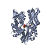
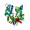
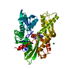
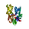
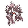
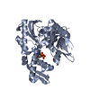
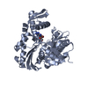
EndoplasmicreticulumchaperoneBiP / 78 kDa glucose-regulated protein / GRP-78 / Binding-immunoglobulin protein / BiP / Heat shock ...78 kDa glucose-regulated protein / GRP-78 / Binding-immunoglobulin protein / BiP / Heat shock protein 70 family protein 5 / HSP70 family protein 5 / Heat shock protein family A member 5 / Immunoglobulin heavy chain-binding protein
Mass: 42155.715 Da / Num. of mol.: 2 / Fragment: UNP residues 26-407 Source method: isolated from a genetically manipulated source Source: (gene. exp.) Homo sapiens (human) / Gene: HSPA5, GRP78 / Plasmid: pET28-MHL / Production host: Escherichia coli (E. coli) References: UniProt: P11021, non-chaperonin molecular chaperone ATPase
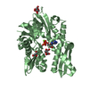
Mass: 18.015 Da / Num. of mol.: 196 / Source method: isolated from a natural source / Formula: H2O
-
Experimental details
-
Experiment
Experiment
Method: X-RAY DIFFRACTION / Number of used crystals: 1
-
Sample preparation
Crystal
Density Matthews: 2.18 Å3/Da / Density % sol: 43.63 % / Description: Plates
Crystal grow
Temperature: 298 K / Method: vapor diffusion, sitting drop / pH: 8.5 Details: Triclinic crystals of apo GRP78 were observed in 0.1 M Tris-HCl, 25% PEG3350, 0.1 M sodium/potassium tartrate. Initial crystals were then microseeded in 25% PEG3350, 0.1 M Tris-HCl, 0.2 M ...Details: Triclinic crystals of apo GRP78 were observed in 0.1 M Tris-HCl, 25% PEG3350, 0.1 M sodium/potassium tartrate. Initial crystals were then microseeded in 25% PEG3350, 0.1 M Tris-HCl, 0.2 M sodium chloride. These new apo monoclinic crystals were soaked with 5 mM 8-bromoadenosine for one week. Temp details: Room temperature
In the structure databanks used in Yorodumi, some data are registered as the other names, "COVID-19 virus" and "2019-nCoV". Here are the details of the virus and the list of structure data.
Jan 31, 2019. EMDB accession codes are about to change! (news from PDBe EMDB page)
EMDB accession codes are about to change! (news from PDBe EMDB page)
The allocation of 4 digits for EMDB accession codes will soon come to an end. Whilst these codes will remain in use, new EMDB accession codes will include an additional digit and will expand incrementally as the available range of codes is exhausted. The current 4-digit format prefixed with “EMD-” (i.e. EMD-XXXX) will advance to a 5-digit format (i.e. EMD-XXXXX), and so on. It is currently estimated that the 4-digit codes will be depleted around Spring 2019, at which point the 5-digit format will come into force.
The EM Navigator/Yorodumi systems omit the EMD- prefix.
Related info.:Q: What is EMD? / ID/Accession-code notation in Yorodumi/EM Navigator
Yorodumi is a browser for structure data from EMDB, PDB, SASBDB, etc.
This page is also the successor to EM Navigator detail page, and also detail information page/front-end page for Omokage search.
The word "yorodu" (or yorozu) is an old Japanese word meaning "ten thousand". "mi" (miru) is to see.
Related info.:EMDB / PDB / SASBDB / Comparison of 3 databanks / Yorodumi Search / Aug 31, 2016. New EM Navigator & Yorodumi / Yorodumi Papers / Jmol/JSmol / Function and homology information / Changes in new EM Navigator and Yorodumi
 Movie
Movie Controller

 Yorodumi
 Open data
 Basic information
 Components
 Keywords
 Function and homology information
 Homo sapiens (human)
 X-RAY DIFFRACTION /
  Authors
 Citation
 Structure visualization
 Downloads & links
 Other downloads
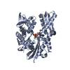


 PDBj
PDBj


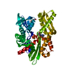
 Assembly
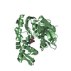


 Mass: 346.137 Da / Num. of mol.: 2 / Source method: obtained synthetically / Formula: C10H12BrN5O4
 Mass: 18.015 Da / Num. of mol.: 196 / Source method: isolated from a natural source / Formula: H2O
 Sample preparation
 / Beamline: 23-ID-D / Wavelength: 1.03327 Å
 Processing