 Movie
Movie Controller

[English] 日本語
 Yorodumi
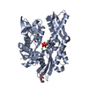
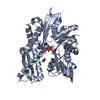
- PDB-3fe1: Crystal structure of the human 70kDa heat shock protein 6 (Hsp70B... -
+ Open data

- Basic information
| Entry | Database: PDB / ID: 3fe1 | ||||||
|---|---|---|---|---|---|---|---|
| Title | Crystal structure of the human 70kDa heat shock protein 6 (Hsp70B') ATPase domain in complex with ADP and inorganic phosphate | ||||||
 Components Components | Heat shock 70 kDa protein 6 | ||||||
 Keywords Keywords | CHAPERONE / mixed beta-sheet / ATP-binding / Nucleotide-binding / Polymorphism / Stress response / Structural Genomics / Structural Genomics Consortium / SGC | ||||||
| Function / homology |  Function and homology information Function and homology informationRegulation of HSF1-mediated heat shock response / response to unfolded protein / heat shock protein binding / protein folding chaperone / centriole / ATP-dependent protein folding chaperone / : / cellular response to heat / protein refolding / secretory granule lumen ...Regulation of HSF1-mediated heat shock response / response to unfolded protein / heat shock protein binding / protein folding chaperone / centriole / ATP-dependent protein folding chaperone / : / cellular response to heat / protein refolding / secretory granule lumen / blood microparticle / ficolin-1-rich granule lumen / Neutrophil degranulation / enzyme binding / ATP hydrolysis activity / extracellular exosome / extracellular region / ATP binding / nucleus / plasma membrane / cytosol / cytoplasm Similarity search - Function | ||||||
| Biological species |  Homo sapiens (human) Homo sapiens (human) | ||||||
| Method |  X-RAY DIFFRACTION / SYNCHROTRON / MOLECULAR REPLACEMENT / molecular replacement / Resolution: 2.2 Å X-RAY DIFFRACTION / SYNCHROTRON / MOLECULAR REPLACEMENT / molecular replacement / Resolution: 2.2 Å | ||||||
 Authors Authors | Wisniewska, M. / Lehtio, L. / Arrowsmith, C.H. / Berglund, H. / Bountra, C. / Collins, R. / Dahlgren, L.G. / Edwards, A.M. / Flodin, S. / Flores, A. ...Wisniewska, M. / Lehtio, L. / Arrowsmith, C.H. / Berglund, H. / Bountra, C. / Collins, R. / Dahlgren, L.G. / Edwards, A.M. / Flodin, S. / Flores, A. / Graslund, S. / Hammarstrom, M. / Johansson, A. / Johansson, I. / Karlberg, T. / Kotenyova, T. / Moche, M. / Nilsson, M.E. / Nordlund, P. / Nyman, T. / Persson, C. / Sagemark, J. / Siponen, M.I. / Thorsell, A.G. / Tresaugues, L. / Van Den Berg, S. / Weigelt, J. / Welin, M. / Wikstrom, M. / Schueler, H. / Structural Genomics Consortium (SGC) | ||||||
 Citation Citation | Journal: Plos One / Year: 2010 Title: Crystal structures of the ATPase domains of four human Hsp70 isoforms: HSPA1L/Hsp70-hom, HSPA2/Hsp70-2, HSPA6/Hsp70B', and HSPA5/BiP/GRP78 Authors: Wisniewska, M. / Karlberg, T. / Lehtio, L. / Johansson, I. / Kotenyova, T. / Moche, M. / Schueler, H. | ||||||
| History |
|
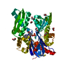
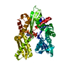
- Structure visualization
| Structure viewer | Molecule: MolmilJmol/JSmol |
|---|
- Downloads & links
-Download
| PDBx/mmCIF format | 3fe1.cif.gz | 240.9 KB | Display | PDBx/mmCIF format |
|---|---|---|---|---|
| PDB format | pdb3fe1.ent.gz | 191.1 KB | Display | PDB format |
| PDBx/mmJSON format | 3fe1.json.gz | Tree view | PDBx/mmJSON format | |
| Others |  Other downloads Other downloads |
-Validation report
| Arichive directory | https://data.pdbj.org/pub/pdb/validation_reports/fe/3fe1ftp://data.pdbj.org/pub/pdb/validation_reports/fe/3fe1 | HTTPS FTP |
|---|
-Related structure data
| Related structure data |  3gdqC  3i33C  3iucC 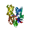 3jxuC  2e88S S: Starting model for refinement C: citing same article ( |
|---|---|
| Similar structure data |


-Links
 PDBj
PDBj


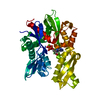
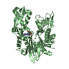
- Assembly
| Deposited unit | 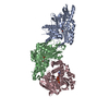
| ||||||||
|---|---|---|---|---|---|---|---|---|---|
| 1 | 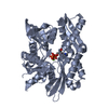
| ||||||||
| 2 | 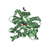
| ||||||||
| 3 | 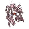
| ||||||||
| Unit cell |
|
-Components
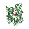
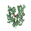
-Protein , 1 types, 3 molecules ABC
| #1: Protein | Mass: 44448.078 Da / Num. of mol.: 3 / Fragment: ATP-ase domain, residues 6-385 Source method: isolated from a genetically manipulated source Source: (gene. exp.) Homo sapiens (human) / Gene: HSPA6, HSP70B' / Plasmid: pNIC-Bsa4 / Production host:  |
|---|
-Non-polymers , 6 types, 344 molecules 


| #2: Chemical |  Mass: 427.201 Da / Num. of mol.: 3 / Source method: obtained synthetically / Formula: C10H15N5O10P2 / Comment: ADP, energy-carrying molecule*YM Mass: 427.201 Da / Num. of mol.: 3 / Source method: obtained synthetically / Formula: C10H15N5O10P2 / Comment: ADP, energy-carrying molecule*YM#3: Chemical |  Mass: 94.971 Da / Num. of mol.: 3 / Source method: obtained synthetically / Formula: PO4 Mass: 94.971 Da / Num. of mol.: 3 / Source method: obtained synthetically / Formula: PO4#4: Chemical |  Mass: 24.305 Da / Num. of mol.: 3 / Source method: obtained synthetically / Formula: Mg Mass: 24.305 Da / Num. of mol.: 3 / Source method: obtained synthetically / Formula: Mg#5: Chemical | ChemComp-CL / |  Mass: 35.453 Da / Num. of mol.: 1 / Source method: obtained synthetically / Formula: Cl Mass: 35.453 Da / Num. of mol.: 1 / Source method: obtained synthetically / Formula: Cl#6: Chemical | ChemComp-PGE / |  Mass: 150.173 Da / Num. of mol.: 1 / Source method: obtained synthetically / Formula: C6H14O4 Mass: 150.173 Da / Num. of mol.: 1 / Source method: obtained synthetically / Formula: C6H14O4#7: Water | ChemComp-HOH / | Mass: 18.015 Da / Num. of mol.: 333 / Source method: isolated from a natural source / Formula: H2O |
|---|
-Experimental details
-Experiment
| Experiment | Method: X-RAY DIFFRACTION / Number of used crystals: 1 |
|---|
- Sample preparation
| Crystal | Density Matthews: 3.37 Å3/Da / Density % sol: 63.55 % |
|---|---|
| Crystal grow | Temperature: 293 K / Method: vapor diffusion, sitting drop / pH: 3.2 Details: 0.1M citric acid, 16% PEG 300, 0.1M di-sodium hydrogen phosphate, pH 3.2, VAPOR DIFFUSION, SITTING DROP, temperature 293K |
-Data collection
| Diffraction | Mean temperature: 100 K |
|---|---|
| Diffraction source | Source: SYNCHROTRON / Site: BESSY  / Beamline: 14.1 / Wavelength: 0.91841 Å / Beamline: 14.1 / Wavelength: 0.91841 Å |
| Detector | Type: MARMOSAIC 225 mm CCD / Detector: CCD / Date: Oct 30, 2008 |
| Radiation | Monochromator: double crystal / Protocol: SINGLE WAVELENGTH / Monochromatic (M) / Laue (L): M / Scattering type: x-ray |
| Radiation wavelength | Wavelength: 0.91841 Å / Relative weight: 1 |
| Reflection | Resolution: 2.2→35 Å / Num. all: 89951 / Num. obs: 88612 / % possible obs: 98.5 % / Redundancy: 3.86 % / Net I/σ(I): 10.54 |
| Reflection shell | Resolution: 2.2→2.26 Å / % possible all: 95 |
-Phasing
| Phasing | Method: molecular replacement |
|---|
- Processing
| Software |
| |||||||||||||||||||||||||||||||||||||||||||||||||||||||||||||||||||||||||||||||||||||
|---|---|---|---|---|---|---|---|---|---|---|---|---|---|---|---|---|---|---|---|---|---|---|---|---|---|---|---|---|---|---|---|---|---|---|---|---|---|---|---|---|---|---|---|---|---|---|---|---|---|---|---|---|---|---|---|---|---|---|---|---|---|---|---|---|---|---|---|---|---|---|---|---|---|---|---|---|---|---|---|---|---|---|---|---|---|---|
| Refinement | Method to determine structure: MOLECULAR REPLACEMENT Starting model: PDB ENTRY 2E88 Resolution: 2.2→33.69 Å / Cor.coef. Fo:Fc: 0.932 / Cor.coef. Fo:Fc free: 0.907 / Occupancy max: 1 / Occupancy min: 0.5 / SU B: 4.428 / SU ML: 0.114 / Cross valid method: THROUGHOUT / σ(F): 0 / ESU R: 0.199 / ESU R Free: 0.177 / Stereochemistry target values: MAXIMUM LIKELIHOOD Details: HYDROGENS HAVE BEEN ADDED IN THE RIDING POSITIONS U VALUES : REFINED INDIVIDUALLY
| |||||||||||||||||||||||||||||||||||||||||||||||||||||||||||||||||||||||||||||||||||||
| Solvent computation | Ion probe radii: 0.8 Å / Shrinkage radii: 0.8 Å / VDW probe radii: 1.2 Å / Solvent model: MASK | |||||||||||||||||||||||||||||||||||||||||||||||||||||||||||||||||||||||||||||||||||||
| Displacement parameters | Biso max: 65.38 Å2 / Biso mean: 25.411 Å2 / Biso min: 6.25 Å2
| |||||||||||||||||||||||||||||||||||||||||||||||||||||||||||||||||||||||||||||||||||||
| Refinement step | Cycle: LAST / Resolution: 2.2→33.69 Å
| |||||||||||||||||||||||||||||||||||||||||||||||||||||||||||||||||||||||||||||||||||||
| Refine LS restraints |
| |||||||||||||||||||||||||||||||||||||||||||||||||||||||||||||||||||||||||||||||||||||
| LS refinement shell | Resolution: 2.2→2.257 Å / Total num. of bins used: 20
|
