 Movie
Movie Controller

[English] 日本語
 Yorodumi
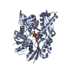
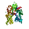
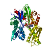
- PDB-3jxu: Crystal structure of the human 70kDa heat shock protein 1A (Hsp70... -
+ Open data

- Basic information
| Entry | Database: PDB / ID: 3jxu | ||||||
|---|---|---|---|---|---|---|---|
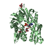
| Title | Crystal structure of the human 70kDa heat shock protein 1A (Hsp70-1) ATPase domain in complex with ADP and inorganic phosphate | ||||||
 Components Components | Heat shock 70 kDa protein 1 | ||||||
 Keywords Keywords | CHAPERONE / helix / Structural Genomics / Structural Genomics Consortium / SGC / ATP-binding / Nucleotide-binding / Phosphoprotein / Polymorphism / Stress response | ||||||
| Function / homology |  Function and homology information Function and homology information: / : / : / : / cellular heat acclimation / negative regulation of inclusion body assembly / Viral RNP Complexes in the Host Cell Nucleus / C3HC4-type RING finger domain binding / positive regulation of nucleotide-binding oligomerization domain containing 2 signaling pathway / positive regulation of microtubule nucleation ...: / : / : / : / cellular heat acclimation / negative regulation of inclusion body assembly / Viral RNP Complexes in the Host Cell Nucleus / C3HC4-type RING finger domain binding / positive regulation of nucleotide-binding oligomerization domain containing 2 signaling pathway / positive regulation of microtubule nucleation / ATP-dependent protein disaggregase activity / misfolded protein binding / negative regulation of mitochondrial outer membrane permeabilization involved in apoptotic signaling pathway / regulation of mitotic spindle assembly / positive regulation of tumor necrosis factor-mediated signaling pathway / aggresome / lysosomal transport / cellular response to steroid hormone stimulus / mRNA catabolic process / regulation of protein ubiquitination / cellular response to unfolded protein / HSF1-dependent transactivation / Regulation of HSF1-mediated heat shock response / response to unfolded protein / Mitochondrial unfolded protein response (UPRmt) / negative regulation of extrinsic apoptotic signaling pathway in absence of ligand / Attenuation phase / negative regulation of endoplasmic reticulum stress-induced intrinsic apoptotic signaling pathway / chaperone-mediated protein complex assembly / transcription regulator inhibitor activity / ATP metabolic process / vesicle-mediated transport / endoplasmic reticulum unfolded protein response / heat shock protein binding / protein folding chaperone / inclusion body / negative regulation of protein ubiquitination / positive regulation of erythrocyte differentiation / HSP90 chaperone cycle for steroid hormone receptors (SHR) in the presence of ligand / positive regulation of RNA splicing / : / positive regulation of interleukin-8 production / ATP-dependent protein folding chaperone / centriole / negative regulation of transforming growth factor beta receptor signaling pathway / AUF1 (hnRNP D0) binds and destabilizes mRNA / negative regulation of cell growth / PKR-mediated signaling / G protein-coupled receptor binding / histone deacetylase binding / disordered domain specific binding / : / transcription corepressor activity / positive regulation of proteasomal ubiquitin-dependent protein catabolic process / cellular response to heat / protein refolding / virus receptor activity / cellular response to oxidative stress / blood microparticle / vesicle / ficolin-1-rich granule lumen / nuclear speck / protein stabilization / cadherin binding / ribonucleoprotein complex / receptor ligand activity / signaling receptor binding / negative regulation of cell population proliferation / focal adhesion / Neutrophil degranulation / ubiquitin protein ligase binding / positive regulation of gene expression / centrosome / negative regulation of apoptotic process / perinuclear region of cytoplasm / enzyme binding / negative regulation of transcription by RNA polymerase II / endoplasmic reticulum / ATP hydrolysis activity / protein-containing complex / mitochondrion / : / RNA binding / extracellular exosome / extracellular region / nucleoplasm / ATP binding / nucleus / plasma membrane / cytoplasm / cytosol Similarity search - Function | ||||||
| Biological species |  Homo sapiens (human) Homo sapiens (human) | ||||||
| Method |  X-RAY DIFFRACTION / MOLECULAR REPLACEMENT / Resolution: 2.14 Å X-RAY DIFFRACTION / MOLECULAR REPLACEMENT / Resolution: 2.14 Å | ||||||
 Authors Authors | Wisniewska, M.M. / Karlberg, T. / Arrowsmith, C.H. / Berglund, H. / Bountra, C. / Collins, R. / Edwards, A.M. / Flodin, S. / Flores, A. / Graslund, S. ...Wisniewska, M.M. / Karlberg, T. / Arrowsmith, C.H. / Berglund, H. / Bountra, C. / Collins, R. / Edwards, A.M. / Flodin, S. / Flores, A. / Graslund, S. / Hammarstrom, M. / Johansson, A. / Johansson, I. / Kallas, A. / Kraulis, P. / Kotenyova, T. / Kotzsch, A. / Markova, N. / Moche, M. / Nielsen, T.K. / Nordlund, P. / Nyman, T. / Persson, C. / Roos, A.K. / Siponen, M.I. / Schutz, P. / Svensson, L. / Thorsell, A.G. / Tresaugues, L. / Van Den Berg, S. / Wahlberg, E. / Weigelt, J. / Welin, M. / Schuler, H. / Structural Genomics Consortium (SGC) | ||||||
 Citation Citation | Journal: Plos One / Year: 2010 Title: Crystal structures of the ATPase domains of four human Hsp70 isoforms: HSPA1L/Hsp70-hom, HSPA2/Hsp70-2, HSPA6/Hsp70B', and HSPA5/BiP/GRP78 Authors: Wisniewska, M. / Karlberg, T. / Lehtio, L. / Johansson, I. / Kotenyova, T. / Moche, M. / Schuler, H. | ||||||
| History |
|
- Structure visualization
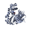
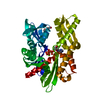
| Structure viewer | Molecule: MolmilJmol/JSmol |
|---|
- Downloads & links
-Download
| PDBx/mmCIF format | 3jxu.cif.gz | 94.7 KB | Display | PDBx/mmCIF format |
|---|---|---|---|---|
| PDB format | pdb3jxu.ent.gz | 69 KB | Display | PDB format |
| PDBx/mmJSON format | 3jxu.json.gz | Tree view | PDBx/mmJSON format | |
| Others |  Other downloads Other downloads |
-Validation report
| Arichive directory | https://data.pdbj.org/pub/pdb/validation_reports/jx/3jxuftp://data.pdbj.org/pub/pdb/validation_reports/jx/3jxu | HTTPS FTP |
|---|
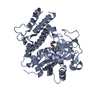
-Related structure data
| Related structure data |  3fe1C  3gdqC  3i33C  3iucC 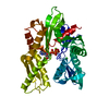 1ba0S C: citing same article ( S: Starting model for refinement |
|---|---|
| Similar structure data |


-Links
 PDBj
PDBj


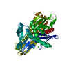
- Assembly
| Deposited unit | 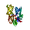
| ||||||||
|---|---|---|---|---|---|---|---|---|---|
| 1 |
| ||||||||
| Unit cell |
|
-Components
| #1: Protein | Mass: 45217.961 Da / Num. of mol.: 1 / Fragment: ATPase domain, UNP resiudes 1-387 Source method: isolated from a genetically manipulated source Source: (gene. exp.) Homo sapiens (human) / Plasmid: PNIC-BSA4 / Production host:  |
|---|---|
| #2: Chemical | ChemComp-ADP /   Mass: 427.201 Da / Num. of mol.: 1 / Source method: obtained synthetically / Formula: C10H15N5O10P2 / Comment: ADP, energy-carrying molecule*YM Mass: 427.201 Da / Num. of mol.: 1 / Source method: obtained synthetically / Formula: C10H15N5O10P2 / Comment: ADP, energy-carrying molecule*YM |
| #3: Chemical | ChemComp-MG /   Mass: 24.305 Da / Num. of mol.: 1 / Source method: obtained synthetically / Formula: Mg Mass: 24.305 Da / Num. of mol.: 1 / Source method: obtained synthetically / Formula: Mg |
| #4: Chemical | ChemComp-PO4 /   Mass: 94.971 Da / Num. of mol.: 1 / Source method: obtained synthetically / Formula: PO4 Mass: 94.971 Da / Num. of mol.: 1 / Source method: obtained synthetically / Formula: PO4 |
| #5: Water | ChemComp-HOH /  Mass: 18.015 Da / Num. of mol.: 202 / Source method: isolated from a natural source / Formula: H2O Mass: 18.015 Da / Num. of mol.: 202 / Source method: isolated from a natural source / Formula: H2O |
-Experimental details
-Experiment
| Experiment | Method: X-RAY DIFFRACTION / Number of used crystals: 1 |
|---|
- Sample preparation
| Crystal | Density Matthews: 2.32 Å3/Da / Density % sol: 47.03 % |
|---|---|
| Crystal grow | Temperature: 277 K / Method: vapor diffusion, sitting drop / pH: 5.5 Details: 0.2M magnesium chloride hexahydrate, 0.1M bis-tris, 25% PEG 3350, pH 5.5, VAPOR DIFFUSION, SITTING DROP, temperature 277K |
-Data collection
| Diffraction source | Source: ROTATING ANODE / Type: OTHER / Wavelength: 1.54166 Å |
|---|---|
| Detector | Type: Bruker Platinum 135 / Detector: CCD / Date: Sep 26, 2008 |
| Radiation | Protocol: SINGLE WAVELENGTH / Monochromatic (M) / Laue (L): M / Scattering type: x-ray |
| Radiation wavelength | Wavelength: 1.54166 Å / Relative weight: 1 |
| Reflection | Resolution: 2.14→26.43 Å / Num. obs: 23808 / % possible obs: 98.3 % / Redundancy: 12.7 % / Rmerge(I) obs: 0.066 |
- Processing
| Software |
| |||||||||||||||||||||||||||||||||||||||||||||||||||||||||||||||||||||||||||||||||||||
|---|---|---|---|---|---|---|---|---|---|---|---|---|---|---|---|---|---|---|---|---|---|---|---|---|---|---|---|---|---|---|---|---|---|---|---|---|---|---|---|---|---|---|---|---|---|---|---|---|---|---|---|---|---|---|---|---|---|---|---|---|---|---|---|---|---|---|---|---|---|---|---|---|---|---|---|---|---|---|---|---|---|---|---|---|---|---|
| Refinement | Method to determine structure: MOLECULAR REPLACEMENT Starting model: PDB ENTRY 1BA0 Resolution: 2.14→10.54 Å / Cor.coef. Fo:Fc: 0.916 / Cor.coef. Fo:Fc free: 0.86 / SU B: 5.788 / SU ML: 0.15 / Cross valid method: THROUGHOUT / ESU R: 0.256 / ESU R Free: 0.215 / Stereochemistry target values: MAXIMUM LIKELIHOOD / Details: HYDROGENS HAVE BEEN ADDED IN THE RIDING POSITIONS
| |||||||||||||||||||||||||||||||||||||||||||||||||||||||||||||||||||||||||||||||||||||
| Solvent computation | Ion probe radii: 0.8 Å / Shrinkage radii: 0.8 Å / VDW probe radii: 1.2 Å / Solvent model: MASK | |||||||||||||||||||||||||||||||||||||||||||||||||||||||||||||||||||||||||||||||||||||
| Displacement parameters | Biso mean: 15.561 Å2
| |||||||||||||||||||||||||||||||||||||||||||||||||||||||||||||||||||||||||||||||||||||
| Refinement step | Cycle: LAST / Resolution: 2.14→10.54 Å
| |||||||||||||||||||||||||||||||||||||||||||||||||||||||||||||||||||||||||||||||||||||
| Refine LS restraints |
| |||||||||||||||||||||||||||||||||||||||||||||||||||||||||||||||||||||||||||||||||||||
| LS refinement shell | Resolution: 2.136→2.189 Å / Total num. of bins used: 20
|
