 Movie
Movie Controller

[English] 日本語
 Yorodumi
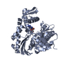
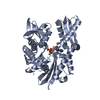
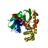
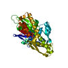
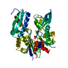
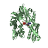
- PDB-3gdq: Crystal structure of the human 70kDa heat shock protein 1-like AT... -
+ Open data

- Basic information
| Entry | Database: PDB / ID: 3gdq | ||||||
|---|---|---|---|---|---|---|---|
| Title | Crystal structure of the human 70kDa heat shock protein 1-like ATPase domain in complex with ADP and inorganic phosphate | ||||||
 Components Components | Heat shock 70 kDa protein 1-like | ||||||
 Keywords Keywords | CHAPERONE / helix / Structural Genomics / Structural Genomics Consortium / SGC / ATP-binding / Nucleotide-binding / TRANSCRIPTION | ||||||
| Function / homology |  Function and homology information Function and homology informationzona pellucida receptor complex / binding of sperm to zona pellucida / : / HSF1-dependent transactivation / Regulation of HSF1-mediated heat shock response / response to unfolded protein / Attenuation phase / heat shock protein binding / protein folding chaperone / HSP90 chaperone cycle for steroid hormone receptors (SHR) in the presence of ligand ...zona pellucida receptor complex / binding of sperm to zona pellucida / : / HSF1-dependent transactivation / Regulation of HSF1-mediated heat shock response / response to unfolded protein / Attenuation phase / heat shock protein binding / protein folding chaperone / HSP90 chaperone cycle for steroid hormone receptors (SHR) in the presence of ligand / ATP-dependent protein folding chaperone / PKR-mediated signaling / : / protein refolding / cell body / blood microparticle / ubiquitin protein ligase binding / ATP hydrolysis activity / nucleoplasm / ATP binding / nucleus / plasma membrane / cytoplasm / cytosol Similarity search - Function | ||||||
| Biological species |  Homo sapiens (human) Homo sapiens (human) | ||||||
| Method |  X-RAY DIFFRACTION / SYNCHROTRON / MOLECULAR REPLACEMENT / Resolution: 1.8 Å X-RAY DIFFRACTION / SYNCHROTRON / MOLECULAR REPLACEMENT / Resolution: 1.8 Å | ||||||
 Authors Authors | Wisniewska, M. / Moche, M. / Arrowsmith, C.H. / Berglund, H. / Bountra, C. / Collins, R. / Dahlgren, L.G. / Edwards, A.M. / Flodin, S. / Flores, A. ...Wisniewska, M. / Moche, M. / Arrowsmith, C.H. / Berglund, H. / Bountra, C. / Collins, R. / Dahlgren, L.G. / Edwards, A.M. / Flodin, S. / Flores, A. / Graslund, S. / Hammarstrom, M. / Johansson, A. / Johansson, I. / Karlberg, T. / Kotenyova, T. / Lehtio, L. / Nilsson, M.E. / Nordlund, P. / Nyman, T. / Persson, C. / Sagemark, J. / Schutz, P. / Siponen, M. / Thorsell, A.G. / Tresaugues, L. / Van Den Berg, S. / Weigelt, J. / Welin, M. / Schueler, H. / Structural Genomics Consortium (SGC) | ||||||
 Citation Citation | Journal: Plos One / Year: 2010 Title: Crystal structures of the ATPase domains of four human Hsp70 isoforms: HSPA1L/Hsp70-hom, HSPA2/Hsp70-2, HSPA6/Hsp70B', and HSPA5/BiP/GRP78 Authors: Wisniewska, M. / Karlberg, T. / Lehtio, L. / Johansson, I. / Kotenyova, T. / Moche, M. / Schuler, H. | ||||||
| History |
|
- Structure visualization
| Structure viewer | Molecule: MolmilJmol/JSmol |
|---|
- Downloads & links
-Download
| PDBx/mmCIF format | 3gdq.cif.gz | 98.2 KB | Display | PDBx/mmCIF format |
|---|---|---|---|---|
| PDB format | pdb3gdq.ent.gz | 71 KB | Display | PDB format |
| PDBx/mmJSON format | 3gdq.json.gz | Tree view | PDBx/mmJSON format | |
| Others |  Other downloads Other downloads |
-Validation report
| Arichive directory | https://data.pdbj.org/pub/pdb/validation_reports/gd/3gdqftp://data.pdbj.org/pub/pdb/validation_reports/gd/3gdq | HTTPS FTP |
|---|
-Related structure data
| Related structure data |  3fe1C  3i33C  3iucC 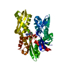 3jxuC 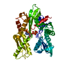 1ba0S S: Starting model for refinement C: citing same article ( |
|---|---|
| Similar structure data |


-Links
 PDBj
PDBj


- Assembly
| Deposited unit | 
| ||||||||
|---|---|---|---|---|---|---|---|---|---|
| 1 |
| ||||||||
| Unit cell |
|
-Components
-Protein , 1 types, 1 molecules A
| #1: Protein | Mass: 44988.910 Da / Num. of mol.: 1 / Fragment: ATPase domain Source method: isolated from a genetically manipulated source Source: (gene. exp.) Homo sapiens (human) / Gene: HSPA1L / Production host:  |
|---|
-Non-polymers , 5 types, 313 molecules 


| #2: Chemical | ChemComp-PO4 /  Mass: 94.971 Da / Num. of mol.: 1 / Source method: obtained synthetically / Formula: PO4 Mass: 94.971 Da / Num. of mol.: 1 / Source method: obtained synthetically / Formula: PO4 |
|---|---|
| #3: Chemical | ChemComp-ADP /  Mass: 427.201 Da / Num. of mol.: 1 / Source method: obtained synthetically / Formula: C10H15N5O10P2 / Comment: ADP, energy-carrying molecule*YM Mass: 427.201 Da / Num. of mol.: 1 / Source method: obtained synthetically / Formula: C10H15N5O10P2 / Comment: ADP, energy-carrying molecule*YM |
| #4: Chemical | ChemComp-GOL /  Mass: 92.094 Da / Num. of mol.: 1 / Source method: obtained synthetically / Formula: C3H8O3 Mass: 92.094 Da / Num. of mol.: 1 / Source method: obtained synthetically / Formula: C3H8O3 |
| #5: Chemical | ChemComp-MN /  Mass: 54.938 Da / Num. of mol.: 1 / Source method: obtained synthetically / Formula: Mn Mass: 54.938 Da / Num. of mol.: 1 / Source method: obtained synthetically / Formula: Mn |
| #6: Water | ChemComp-HOH / Mass: 18.015 Da / Num. of mol.: 309 / Source method: isolated from a natural source / Formula: H2O |
-Experimental details
-Experiment
| Experiment | Method: X-RAY DIFFRACTION / Number of used crystals: 1 |
|---|
- Sample preparation
| Crystal | Density Matthews: 2.68 Å3/Da / Density % sol: 54.17 % |
|---|---|
| Crystal grow | Temperature: 277 K / Method: vapor diffusion, sitting drop / pH: 8.5 Details: 26% PEG monomethyl ether 2000, 0.1M Tris, 0.2M trimethylamine n-oxide, pH 8.5, VAPOR DIFFUSION, SITTING DROP, temperature 277K |
-Data collection
| Diffraction | Mean temperature: 77 K |
|---|---|
| Diffraction source | Source: SYNCHROTRON / Site: Diamond  / Beamline: I03 / Wavelength: 0.98 Å / Beamline: I03 / Wavelength: 0.98 Å |
| Detector | Type: ADSC QUANTUM 315 / Detector: CCD / Date: Dec 12, 2008 |
| Radiation | Monochromator: DOUBLE CRYSTAL, SI(111) / Protocol: SINGLE WAVELENGTH / Monochromatic (M) / Laue (L): M / Scattering type: x-ray |
| Radiation wavelength | Wavelength: 0.98 Å / Relative weight: 1 |
| Reflection | Resolution: 1.8→25 Å / Num. all: 45423 / Num. obs: 45400 |
| Reflection shell | Resolution: 1.8→1.85 Å |
- Processing
| Software |
| |||||||||||||||||||||||||||||||||||||||||||||||||||||||||||||||||||||||||||||||||||||
|---|---|---|---|---|---|---|---|---|---|---|---|---|---|---|---|---|---|---|---|---|---|---|---|---|---|---|---|---|---|---|---|---|---|---|---|---|---|---|---|---|---|---|---|---|---|---|---|---|---|---|---|---|---|---|---|---|---|---|---|---|---|---|---|---|---|---|---|---|---|---|---|---|---|---|---|---|---|---|---|---|---|---|---|---|---|---|
| Refinement | Method to determine structure: MOLECULAR REPLACEMENT Starting model: PDB entry 1BA0 Resolution: 1.8→23.83 Å / Cor.coef. Fo:Fc: 0.957 / Cor.coef. Fo:Fc free: 0.94 / SU B: 1.898 / SU ML: 0.061 / Cross valid method: THROUGHOUT / ESU R: 0.104 / ESU R Free: 0.103 / Stereochemistry target values: MAXIMUM LIKELIHOOD / Details: HYDROGENS HAVE BEEN ADDED IN THE RIDING POSITIONS
| |||||||||||||||||||||||||||||||||||||||||||||||||||||||||||||||||||||||||||||||||||||
| Solvent computation | Ion probe radii: 0.8 Å / Shrinkage radii: 0.8 Å / VDW probe radii: 1.2 Å / Solvent model: MASK | |||||||||||||||||||||||||||||||||||||||||||||||||||||||||||||||||||||||||||||||||||||
| Displacement parameters | Biso mean: 15.614 Å2
| |||||||||||||||||||||||||||||||||||||||||||||||||||||||||||||||||||||||||||||||||||||
| Refinement step | Cycle: LAST / Resolution: 1.8→23.83 Å
| |||||||||||||||||||||||||||||||||||||||||||||||||||||||||||||||||||||||||||||||||||||
| Refine LS restraints |
| |||||||||||||||||||||||||||||||||||||||||||||||||||||||||||||||||||||||||||||||||||||
| LS refinement shell | Resolution: 1.8→1.85 Å / Total num. of bins used: 20
|
