 Movie
Movie Controller

[English] 日本語
 Yorodumi
- PDB-5o74: Crystal structure of human Rab1b covalently bound to the GEF doma... -
+ Open data

- Basic information
| Entry | Database: PDB / ID: 5o74 | |||||||||||||||
|---|---|---|---|---|---|---|---|---|---|---|---|---|---|---|---|---|
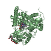
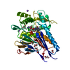
| Title | Crystal structure of human Rab1b covalently bound to the GEF domain of DrrA/SidM from Legionella pneumophila in the presence of GDP | |||||||||||||||
 Components Components |
| |||||||||||||||
 Keywords Keywords | HYDROLASE / Rab1b / DrrA / exchange factor / Legionella pneumophila | |||||||||||||||
| Function / homology |  Function and homology information Function and homology information: / protein guanylylation / positive regulation of glycoprotein metabolic process / protein adenylylation / AMPylase activity / protein adenylyltransferase / phagophore assembly site membrane / RAB geranylgeranylation / regulation of autophagosome assembly / RAB GEFs exchange GTP for GDP on RABs ...: / protein guanylylation / positive regulation of glycoprotein metabolic process / protein adenylylation / AMPylase activity / protein adenylyltransferase / phagophore assembly site membrane / RAB geranylgeranylation / regulation of autophagosome assembly / RAB GEFs exchange GTP for GDP on RABs / regulation of GTPase activity / Golgi Cisternae Pericentriolar Stack Reorganization / phosphatidylinositol-4-phosphate binding / host cell cytoplasmic vesicle / COPII-mediated vesicle transport / COPI-dependent Golgi-to-ER retrograde traffic / virion assembly / Golgi organization / endoplasmic reticulum to Golgi vesicle-mediated transport / autophagosome assembly / transport vesicle / COPI-mediated anterograde transport / endoplasmic reticulum-Golgi intermediate compartment membrane / endomembrane system / guanyl-nucleotide exchange factor activity / small monomeric GTPase / intracellular protein transport / host cell cytoplasmic vesicle membrane / small GTPase binding / protein guanylyltransferase activity / G protein activity / Golgi membrane / GTPase activity / endoplasmic reticulum membrane / GTP binding / perinuclear region of cytoplasm / Golgi apparatus / extracellular exosome / extracellular region / ATP binding / cytosol Similarity search - Function | |||||||||||||||
| Biological species |   Legionella pneumophila (bacteria) Legionella pneumophila (bacteria) Homo sapiens (human) Homo sapiens (human) | |||||||||||||||
| Method |  X-RAY DIFFRACTION / SYNCHROTRON / MOLECULAR REPLACEMENT / Resolution: 2.5 Å X-RAY DIFFRACTION / SYNCHROTRON / MOLECULAR REPLACEMENT / Resolution: 2.5 Å | |||||||||||||||
 Authors Authors | Cigler, M. / Mueller, T. / Horn-Ghetko, D. / von Wrisberg, M.K. / Fottner, M. / Goody, R.S. / Itzen, A. / Mueller, M.P. / Lang, K. | |||||||||||||||
| Funding support |  Germany, 4items Germany, 4items
| |||||||||||||||
 Citation Citation | Journal: Angew. Chem. Int. Ed. Engl. / Year: 2017 Title: Proximity-Triggered Covalent Stabilization of Low-Affinity Protein Complexes In Vitro and In Vivo. Authors: Cigler, M. / Muller, T.G. / Horn-Ghetko, D. / von Wrisberg, M.K. / Fottner, M. / Goody, R.S. / Itzen, A. / Muller, M.P. / Lang, K. | |||||||||||||||
| History |
|
- Structure visualization
| Structure viewer | Molecule: MolmilJmol/JSmol |
|---|
- Downloads & links
-Download
| PDBx/mmCIF format | 5o74.cif.gz | 1.2 MB | Display | PDBx/mmCIF format |
|---|---|---|---|---|
| PDB format | pdb5o74.ent.gz | 1023.9 KB | Display | PDB format |
| PDBx/mmJSON format | 5o74.json.gz | Tree view | PDBx/mmJSON format | |
| Others |  Other downloads Other downloads |
-Validation report
| Arichive directory | https://data.pdbj.org/pub/pdb/validation_reports/o7/5o74ftp://data.pdbj.org/pub/pdb/validation_reports/o7/5o74 | HTTPS FTP |
|---|
-Related structure data
| Related structure data | 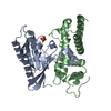 3jzaS S: Starting model for refinement |
|---|---|
| Similar structure data |


-Links
 PDBj
PDBj


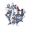
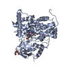
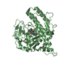
- Assembly
| Deposited unit | 
| ||||||||
|---|---|---|---|---|---|---|---|---|---|
| 1 | 
| ||||||||
| 2 | 
| ||||||||
| 3 | 
| ||||||||
| 4 | 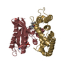
| ||||||||
| 5 | 
| ||||||||
| 6 | 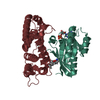
| ||||||||
| Unit cell |
|
-Components
| #1: Protein | Mass: 22001.195 Da / Num. of mol.: 6 / Mutation: D512C Source method: isolated from a genetically manipulated source Source: (gene. exp.) Legionella pneumophila (bacteria) / Gene: drrA, sidM / Production host: References: UniProt: Q29ST3, Transferases; Transferring phosphorus-containing groups; Nucleotidyltransferases #2: Protein | Mass: 20605.148 Da / Num. of mol.: 6 Source method: isolated from a genetically manipulated source Source: (gene. exp.) Homo sapiens (human) / Gene: RAB1B / Production host: #3: Chemical | ChemComp-GDP /   Type: RNA linking / Mass: 443.201 Da / Num. of mol.: 6 Type: RNA linking / Mass: 443.201 Da / Num. of mol.: 6Source method: isolated from a genetically manipulated source Formula: C10H15N5O11P2 / Source: (gene. exp.) Homo sapiens (human) / Gene: RAB1B / Production host: |
|---|
-Experimental details
-Experiment
| Experiment | Method: X-RAY DIFFRACTION / Number of used crystals: 1 |
|---|
- Sample preparation
| Crystal | Density Matthews: 2.27 Å3/Da / Density % sol: 45.73 % |
|---|---|
| Crystal grow | Temperature: 293 K / Method: vapor diffusion, hanging drop / pH: 5.5 Details: 19% PEG 600, 100mM NaCitrate pH 5.5, protein concentration 22mg/ml |
-Data collection
| Diffraction | Mean temperature: 100 K |
|---|---|
| Diffraction source | Source: SYNCHROTRON / Site: SLS  / Beamline: X10SA / Wavelength: 1.00005 Å / Beamline: X10SA / Wavelength: 1.00005 Å |
| Detector | Type: DECTRIS PILATUS 6M / Detector: PIXEL / Date: Feb 26, 2016 |
| Radiation | Monochromator: Si(111) / Protocol: SINGLE WAVELENGTH / Monochromatic (M) / Laue (L): M / Scattering type: x-ray |
| Radiation wavelength | Wavelength: 1.00005 Å / Relative weight: 1 |
| Reflection | Resolution: 2.5→46.525 Å / Num. obs: 72360 / % possible obs: 97.8 % / Redundancy: 3.5 % / Rsym value: 0.092 / Net I/σ(I): 10.2 |
| Reflection shell | Resolution: 2.5→2.6 Å / Redundancy: 3.6 % / Mean I/σ(I) obs: 2.5 / Num. unique obs: 7978 / Rsym value: 0.596 / % possible all: 97.5 |
- Processing
| Software |
| |||||||||||||||||||||||||||||||||||||||||||||||||||||||||||||||||||||||||||||||||||||||||||||||||||||||||||||||||||||||||||||||||||||||||||||||||||||||||||||||||||||||||||||||||||||||||||||
|---|---|---|---|---|---|---|---|---|---|---|---|---|---|---|---|---|---|---|---|---|---|---|---|---|---|---|---|---|---|---|---|---|---|---|---|---|---|---|---|---|---|---|---|---|---|---|---|---|---|---|---|---|---|---|---|---|---|---|---|---|---|---|---|---|---|---|---|---|---|---|---|---|---|---|---|---|---|---|---|---|---|---|---|---|---|---|---|---|---|---|---|---|---|---|---|---|---|---|---|---|---|---|---|---|---|---|---|---|---|---|---|---|---|---|---|---|---|---|---|---|---|---|---|---|---|---|---|---|---|---|---|---|---|---|---|---|---|---|---|---|---|---|---|---|---|---|---|---|---|---|---|---|---|---|---|---|---|---|---|---|---|---|---|---|---|---|---|---|---|---|---|---|---|---|---|---|---|---|---|---|---|---|---|---|---|---|---|---|---|---|
| Refinement | Method to determine structure: MOLECULAR REPLACEMENT Starting model: 3jza Resolution: 2.5→46.525 Å / SU ML: 0.4 / Cross valid method: FREE R-VALUE / σ(F): 2 / Phase error: 32.49
| |||||||||||||||||||||||||||||||||||||||||||||||||||||||||||||||||||||||||||||||||||||||||||||||||||||||||||||||||||||||||||||||||||||||||||||||||||||||||||||||||||||||||||||||||||||||||||||
| Solvent computation | Shrinkage radii: 0.9 Å / VDW probe radii: 1.11 Å | |||||||||||||||||||||||||||||||||||||||||||||||||||||||||||||||||||||||||||||||||||||||||||||||||||||||||||||||||||||||||||||||||||||||||||||||||||||||||||||||||||||||||||||||||||||||||||||
| Refinement step | Cycle: LAST / Resolution: 2.5→46.525 Å
| |||||||||||||||||||||||||||||||||||||||||||||||||||||||||||||||||||||||||||||||||||||||||||||||||||||||||||||||||||||||||||||||||||||||||||||||||||||||||||||||||||||||||||||||||||||||||||||
| Refine LS restraints |
| |||||||||||||||||||||||||||||||||||||||||||||||||||||||||||||||||||||||||||||||||||||||||||||||||||||||||||||||||||||||||||||||||||||||||||||||||||||||||||||||||||||||||||||||||||||||||||||
| LS refinement shell |
| |||||||||||||||||||||||||||||||||||||||||||||||||||||||||||||||||||||||||||||||||||||||||||||||||||||||||||||||||||||||||||||||||||||||||||||||||||||||||||||||||||||||||||||||||||||||||||||
| Refinement TLS params. | Method: refined / Origin x: 18.2404 Å / Origin y: -8.8812 Å / Origin z: -48.605 Å
| |||||||||||||||||||||||||||||||||||||||||||||||||||||||||||||||||||||||||||||||||||||||||||||||||||||||||||||||||||||||||||||||||||||||||||||||||||||||||||||||||||||||||||||||||||||||||||||
| Refinement TLS group | Selection details: all |
