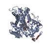
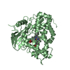
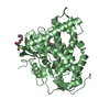
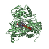
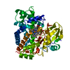
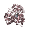
登録情報 データベース : PDB / ID : 4fyzタイトル Crystal Structure of Nitrosyl Cytochrome P450cin P450cin キーワード / / / / 機能・相同性 分子機能 ドメイン・相同性 構成要素
/ / / / / / / / / / / / / / / 生物種 Citrobacter braakii (バクテリア)手法 / / 解像度 : 2.32 Å データ登録者 Madrona, Y. / Tripathi, S.M. / Li, H. / Poulos, T.L. ジャーナル : Biochemistry / 年 : 2012タイトル : Crystal structures of substrate-free and nitrosyl cytochrome p450cin: implications for o(2) activation.著者 : Madrona, Y. / Tripathi, S. / Li, H. / Poulos, T.L. 履歴 登録 2012年7月5日 登録サイト / 処理サイト 改定 1.0 2012年7月25日 Provider / タイプ 改定 1.1 2012年8月1日 Group 改定 1.2 2012年9月5日 Group 改定 1.3 2023年9月13日 Group Data collection / Database references ... Data collection / Database references / Derived calculations / Refinement description カテゴリ chem_comp_atom / chem_comp_bond ... chem_comp_atom / chem_comp_bond / database_2 / pdbx_initial_refinement_model / pdbx_struct_conn_angle / struct_conn / struct_ref_seq_dif / struct_site Item _database_2.pdbx_DOI / _database_2.pdbx_database_accession ... _database_2.pdbx_DOI / _database_2.pdbx_database_accession / _pdbx_struct_conn_angle.ptnr1_auth_asym_id / _pdbx_struct_conn_angle.ptnr1_label_asym_id / _pdbx_struct_conn_angle.ptnr2_auth_asym_id / _pdbx_struct_conn_angle.ptnr2_label_asym_id / _pdbx_struct_conn_angle.ptnr3_auth_asym_id / _pdbx_struct_conn_angle.ptnr3_label_asym_id / _pdbx_struct_conn_angle.value / _struct_conn.pdbx_dist_value / _struct_conn.ptnr1_auth_asym_id / _struct_conn.ptnr1_label_asym_id / _struct_conn.ptnr2_auth_asym_id / _struct_conn.ptnr2_label_asym_id / _struct_conn.ptnr2_label_atom_id / _struct_ref_seq_dif.details / _struct_site.pdbx_auth_asym_id / _struct_site.pdbx_auth_comp_id / _struct_site.pdbx_auth_seq_id
すべて表示 表示を減らす
 ムービー
ムービー コントローラー


 データを開く
 基本情報
基本情報 要素
要素 キーワード
キーワード 機能・相同性情報
 Citrobacter braakii (バクテリア)
 X線回折 /
  データ登録者
 引用
引用 構造の表示
構造の表示 ダウンロードとリンク
 その他のダウンロード
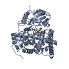


 PDBj
PDBj


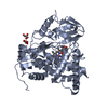
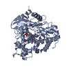
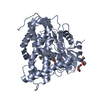
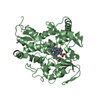
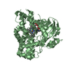
 集合体
集合体
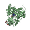


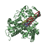
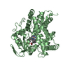
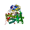
 分子量: 616.487 Da / 分子数: 2 / 由来タイプ: 合成 / 式: C34H32FeN4O4
 分子量: 154.249 Da / 分子数: 2 / 由来タイプ: 合成 / 式: C10H18O
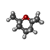
 分子量: 30.006 Da / 分子数: 2 / 由来タイプ: 合成 / 式: NO
 分子量: 194.226 Da / 分子数: 2 / 由来タイプ: 合成 / 式: C8H18O5 / コメント: 沈殿剤*YM
 分子量: 106.120 Da / 分子数: 2 / 由来タイプ: 合成 / 式: C4H10O3
 試料調製
試料調製 解析
解析