 Movie
Movie Controller


+ Open data

- Basic information
| Entry | Database: PDB / ID: 3be0 | ||||||
|---|---|---|---|---|---|---|---|
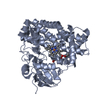
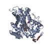
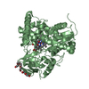
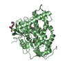
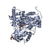
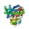
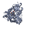
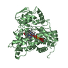
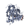
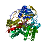
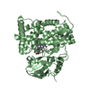
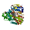
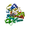
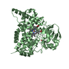
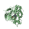
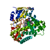
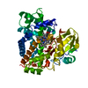
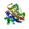
| Title | The Role of Asn 242 in P450cin | ||||||
 Components Components | P450cin | ||||||
 Keywords Keywords | UNKNOWN FUNCTION / Protein substrate complex | ||||||
| Function / homology |  Function and homology information Function and homology information1,8-cineole 2-endo-monooxygenase / carbazole catabolic process / oxidoreductase activity, acting on paired donors, with incorporation or reduction of molecular oxygen, NAD(P)H as one donor, and incorporation of one atom of oxygen / iron ion binding / heme binding Similarity search - Function | ||||||
| Biological species |  Citrobacter braakii (bacteria) Citrobacter braakii (bacteria) | ||||||
| Method |  X-RAY DIFFRACTION / SYNCHROTRON / MOLECULAR REPLACEMENT / Resolution: 3.05 Å X-RAY DIFFRACTION / SYNCHROTRON / MOLECULAR REPLACEMENT / Resolution: 3.05 Å | ||||||
 Authors Authors | Meharenna, Y.T. / Poulos, T.L. | ||||||
 Citation Citation | Journal: J.Biol.Chem. / Year: 2008 Title: The critical role of substrate-protein hydrogen bonding in the control of regioselective hydroxylation in p450cin Authors: Meharenna, Y.T. / Slessor, K.E. / Cavaignac, S.M. / Poulos, T.L. / De Voss, J.J. | ||||||
| History |
|
- Structure visualization
| Structure viewer | Molecule: MolmilJmol/JSmol |
|---|
- Downloads & links
-Download
| PDBx/mmCIF format | 3be0.cif.gz | 171.8 KB | Display | PDBx/mmCIF format |
|---|---|---|---|---|
| PDB format | pdb3be0.ent.gz | 135.3 KB | Display | PDB format |
| PDBx/mmJSON format | 3be0.json.gz | Tree view | PDBx/mmJSON format | |
| Others |  Other downloads Other downloads |
-Validation report
| Arichive directory | https://data.pdbj.org/pub/pdb/validation_reports/be/3be0ftp://data.pdbj.org/pub/pdb/validation_reports/be/3be0 | HTTPS FTP |
|---|
-Related structure data
| Related structure data | 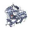 3bdzC  1t2bS S: Starting model for refinement C: citing same article ( |
|---|---|
| Similar structure data |

-Links
 PDBj
PDBj
- Assembly
| Deposited unit | 
| ||||||||
|---|---|---|---|---|---|---|---|---|---|
| 1 | 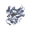
| ||||||||
| 2 | 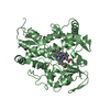
| ||||||||
| Unit cell |
|
-Components
| #1: Protein | Mass: 44613.660 Da / Num. of mol.: 2 / Fragment: UNP residues 8-404 / Mutation: N242A Source method: isolated from a genetically manipulated source Source: (gene. exp.) Citrobacter braakii (bacteria) / Gene: cinA / Plasmid: PCWORI / Production host: #2: Chemical |   Mass: 616.487 Da / Num. of mol.: 2 / Source method: obtained synthetically / Formula: C34H32FeN4O4 Mass: 616.487 Da / Num. of mol.: 2 / Source method: obtained synthetically / Formula: C34H32FeN4O4#3: Chemical |   Mass: 154.249 Da / Num. of mol.: 2 / Source method: obtained synthetically / Formula: C10H18O Mass: 154.249 Da / Num. of mol.: 2 / Source method: obtained synthetically / Formula: C10H18O#4: Water | ChemComp-HOH / |  Mass: 18.015 Da / Num. of mol.: 89 / Source method: isolated from a natural source / Formula: H2O Mass: 18.015 Da / Num. of mol.: 89 / Source method: isolated from a natural source / Formula: H2OHas protein modification | N | |
|---|
-Experimental details
-Experiment
| Experiment | Method: X-RAY DIFFRACTION / Number of used crystals: 1 |
|---|
- Sample preparation
| Crystal | Density Matthews: 2.93 Å3/Da / Density % sol: 58 % |
|---|---|
| Crystal grow | Temperature: 298 K / Method: vapor diffusion, sitting drop / pH: 8 Details: 0.8M sodium malonate, pH 8.0, VAPOR DIFFUSION, SITTING DROP, temperature 298K |
-Data collection
| Diffraction | Mean temperature: 100 K |
|---|---|
| Diffraction source | Source: SYNCHROTRON / Site: ALS  / Beamline: 5.0.2 / Wavelength: 1 Å / Beamline: 5.0.2 / Wavelength: 1 Å |
| Detector | Type: ADSC QUANTUM 315 / Detector: CCD / Date: Dec 2, 2006 / Details: mirrors |
| Radiation | Monochromator: Double crystal / Protocol: SINGLE WAVELENGTH / Monochromatic (M) / Laue (L): M / Scattering type: x-ray |
| Radiation wavelength | Wavelength: 1 Å / Relative weight: 1 |
| Reflection | Resolution: 3.05→50 Å / Num. obs: 17982 / % possible obs: 91.1 % / Observed criterion σ(F): 0 / Observed criterion σ(I): -3 / Redundancy: 6.1 % / Rmerge(I) obs: 0.148 / Rsym value: 0.148 |
| Reflection shell | Resolution: 3.05→3.1 Å / Redundancy: 6.1 % / % possible all: 91.1 |
- Processing
| Software |
| ||||||||||||||||||||
|---|---|---|---|---|---|---|---|---|---|---|---|---|---|---|---|---|---|---|---|---|---|
| Refinement | Method to determine structure: MOLECULAR REPLACEMENT Starting model: PDB ENTRY 1T2B Resolution: 3.05→47.95 Å / Rfactor Rfree error: 0.008 / Data cutoff high absF: 1564056.45 / Data cutoff low absF: 0 / Isotropic thermal model: RESTRAINED / Cross valid method: THROUGHOUT / σ(F): 0 / Stereochemistry target values: Engh & Huber / Details: BULK SOLVENT MODEL USED
| ||||||||||||||||||||
| Solvent computation | Solvent model: FLAT MODEL / Bsol: 51.3266 Å2 / ksol: 0.35 e/Å3 | ||||||||||||||||||||
| Displacement parameters | Biso mean: 59.8 Å2
| ||||||||||||||||||||
| Refine analyze |
| ||||||||||||||||||||
| Refinement step | Cycle: LAST / Resolution: 3.05→47.95 Å
| ||||||||||||||||||||
| Refine LS restraints |
| ||||||||||||||||||||
| LS refinement shell | Resolution: 3.05→3.1 Å / Rfactor Rfree error: 0.034 / Total num. of bins used: 10
| ||||||||||||||||||||
| Xplor file |
|
