 Movie
Movie Controller

[English] 日本語
 Yorodumi
- PDB-3tih: Crystal structure of unliganded HIV-1 clade C strain ZM109F.PB4 g... -
+ Open data

- Basic information
| Entry | Database: PDB / ID: 3tih | ||||||
|---|---|---|---|---|---|---|---|
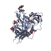
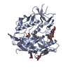
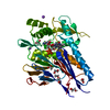
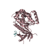
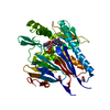
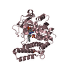
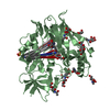
| Title | Crystal structure of unliganded HIV-1 clade C strain ZM109F.PB4 gp120 core | ||||||
 Components Components | HIV-1 clade C ZM109F.PB4 gp120 | ||||||
 Keywords Keywords | VIRAL PROTEIN / HIV-1 gp120 / unliganded / clade C ZM109F.PB4 | ||||||
| Function / homology | HIV Envelope Protein Gp120; Chain G / Human immunodeficiency virus 1, Gp160, envelope glycoprotein / Beta Complex / Mainly Beta Function and homology information Function and homology information | ||||||
| Biological species |   Human immunodeficiency virus 1 Human immunodeficiency virus 1 | ||||||
| Method |  X-RAY DIFFRACTION / SYNCHROTRON / MOLECULAR REPLACEMENT / Resolution: 4 Å X-RAY DIFFRACTION / SYNCHROTRON / MOLECULAR REPLACEMENT / Resolution: 4 Å | ||||||
 Authors Authors | Kwon, Y.D. / Kwong, P.D. | ||||||
 Citation Citation | Journal: Proc.Natl.Acad.Sci.USA / Year: 2012 Title: Unliganded HIV-1 gp120 core structures assume the CD4-bound conformation with regulation by quaternary interactions and variable loops. Authors: Kwon, Y.D. / Finzi, A. / Wu, X. / Dogo-Isonagie, C. / Lee, L.K. / Moore, L.R. / Schmidt, S.D. / Stuckey, J. / Yang, Y. / Zhou, T. / Zhu, J. / Vicic, D.A. / Debnath, A.K. / Shapiro, L. / ...Authors: Kwon, Y.D. / Finzi, A. / Wu, X. / Dogo-Isonagie, C. / Lee, L.K. / Moore, L.R. / Schmidt, S.D. / Stuckey, J. / Yang, Y. / Zhou, T. / Zhu, J. / Vicic, D.A. / Debnath, A.K. / Shapiro, L. / Bewley, C.A. / Mascola, J.R. / Sodroski, J.G. / Kwong, P.D. | ||||||
| History |
|
- Structure visualization
| Structure viewer | Molecule: MolmilJmol/JSmol |
|---|
- Downloads & links
-Download
| PDBx/mmCIF format | 3tih.cif.gz | 261.2 KB | Display | PDBx/mmCIF format |
|---|---|---|---|---|
| PDB format | pdb3tih.ent.gz | 213.2 KB | Display | PDB format |
| PDBx/mmJSON format | 3tih.json.gz | Tree view | PDBx/mmJSON format | |
| Others |  Other downloads Other downloads |
-Validation report
| Arichive directory | https://data.pdbj.org/pub/pdb/validation_reports/ti/3tihftp://data.pdbj.org/pub/pdb/validation_reports/ti/3tih | HTTPS FTP |
|---|
-Related structure data


-Links
 PDBj
PDBj- Assembly
| Deposited unit | 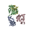
| ||||||||
|---|---|---|---|---|---|---|---|---|---|
| 1 | 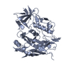
| ||||||||
| 2 | 
| ||||||||
| 3 | 
| ||||||||
| 4 | 
| ||||||||
| Unit cell |
|
-Components
| #1: Protein | Mass: 38547.570 Da / Num. of mol.: 4 Source method: isolated from a genetically manipulated source Source: (gene. exp.) Human immunodeficiency virus 1 / Gene: HIV-1 env / Plasmid: pVRC8400 / Cell line (production host): HEK 293 / Production host:  Homo sapiens (human) Homo sapiens (human)Has protein modification | Y | |
|---|
-Experimental details
-Experiment
| Experiment | Method: X-RAY DIFFRACTION / Number of used crystals: 1 |
|---|
- Sample preparation
| Crystal | Density Matthews: 2.96 Å3/Da / Density % sol: 58.45 % |
|---|---|
| Crystal grow | Temperature: 293 K / Method: vapor diffusion, hanging drop / pH: 7.5 Details: 10% PEG 3350, 10% iso-propanol, 0.2 M Ammonium citrate, VAPOR DIFFUSION, HANGING DROP, temperature 293K, pH 7.5 |
-Data collection
| Diffraction | Mean temperature: 100 K | |||||||||||||||||||||||||||||||||||||||||||||||||||||||||||||||||||||||||||||
|---|---|---|---|---|---|---|---|---|---|---|---|---|---|---|---|---|---|---|---|---|---|---|---|---|---|---|---|---|---|---|---|---|---|---|---|---|---|---|---|---|---|---|---|---|---|---|---|---|---|---|---|---|---|---|---|---|---|---|---|---|---|---|---|---|---|---|---|---|---|---|---|---|---|---|---|---|---|---|
| Diffraction source | Source: SYNCHROTRON / Site: APS  / Beamline: 22-ID / Wavelength: 1 Å / Beamline: 22-ID / Wavelength: 1 Å | |||||||||||||||||||||||||||||||||||||||||||||||||||||||||||||||||||||||||||||
| Detector | Type: MAR scanner 300 mm plate / Detector: IMAGE PLATE / Date: Oct 14, 2010 | |||||||||||||||||||||||||||||||||||||||||||||||||||||||||||||||||||||||||||||
| Radiation | Protocol: SINGLE WAVELENGTH / Monochromatic (M) / Laue (L): M / Scattering type: x-ray | |||||||||||||||||||||||||||||||||||||||||||||||||||||||||||||||||||||||||||||
| Radiation wavelength | Wavelength: 1 Å / Relative weight: 1 | |||||||||||||||||||||||||||||||||||||||||||||||||||||||||||||||||||||||||||||
| Reflection | Resolution: 4→50 Å / Num. all: 15241 / Num. obs: 12925 / % possible obs: 84.8 % / Observed criterion σ(F): 0 / Observed criterion σ(I): 0 / Redundancy: 3.3 % / Rmerge(I) obs: 0.151 / Rsym value: 0.114 / Net I/σ(I): 6.9 | |||||||||||||||||||||||||||||||||||||||||||||||||||||||||||||||||||||||||||||
| Reflection shell |
|
- Processing
| Software |
| |||||||||||||||||||||||||||||||||||
|---|---|---|---|---|---|---|---|---|---|---|---|---|---|---|---|---|---|---|---|---|---|---|---|---|---|---|---|---|---|---|---|---|---|---|---|---|
| Refinement | Method to determine structure: MOLECULAR REPLACEMENT / Resolution: 4→46.695 Å / Occupancy max: 1 / Occupancy min: 1 / SU ML: 1.23 / σ(F): 1.34 / Phase error: 36.58 / Stereochemistry target values: ML
| |||||||||||||||||||||||||||||||||||
| Solvent computation | Shrinkage radii: 0.86 Å / VDW probe radii: 1.1 Å / Solvent model: FLAT BULK SOLVENT MODEL / Bsol: 158.036 Å2 / ksol: 0.33 e/Å3 | |||||||||||||||||||||||||||||||||||
| Displacement parameters |
| |||||||||||||||||||||||||||||||||||
| Refinement step | Cycle: LAST / Resolution: 4→46.695 Å
| |||||||||||||||||||||||||||||||||||
| Refine LS restraints |
| |||||||||||||||||||||||||||||||||||
| LS refinement shell |
|
