 Movie
Movie Controller

[English] 日本語
 Yorodumi
- PDB-6gmg: Structure of a glutamine donor mimicking inhibitory peptide shape... -
+ Open data

- Basic information
| Entry | Database: PDB / ID: 6gmg | ||||||
|---|---|---|---|---|---|---|---|
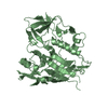
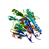
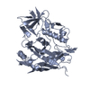
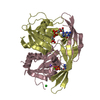
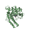
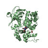
| Title | Structure of a glutamine donor mimicking inhibitory peptide shaped by the catalytic cleft of microbial transglutaminase | ||||||
 Components Components |
| ||||||
 Keywords Keywords | TRANSFERASE / transglutaminase / Streptomyces mobaraensis / peptidic inhibitors / enzyme peptide interaction | ||||||
| Function / homology |  Function and homology information Function and homology informationnegative regulation of peptidase activity / protein-glutamine gamma-glutamyltransferase activity / cysteine-type endopeptidase inhibitor activity / serine-type endopeptidase inhibitor activity / : Similarity search - Function | ||||||
| Biological species |  Streptomyces mobaraensis NBRC 13819 = DSM 40847 (bacteria)Streptomyces mobaraensis (bacteria) Streptomyces mobaraensis NBRC 13819 = DSM 40847 (bacteria)Streptomyces mobaraensis (bacteria) | ||||||
| Method |  X-RAY DIFFRACTION / SYNCHROTRON / MOLECULAR REPLACEMENT / molecular replacement / Resolution: 2.25 Å X-RAY DIFFRACTION / SYNCHROTRON / MOLECULAR REPLACEMENT / molecular replacement / Resolution: 2.25 Å | ||||||
 Authors Authors | Schmelz, S. / Juettner, N.E. / Fuchsbauer, H.L. / Scrima, A. | ||||||
 Citation Citation | Journal: FEBS J. / Year: 2018 Title: Structure of a glutamine donor mimicking inhibitory peptide shaped by the catalytic cleft of microbial transglutaminase. Authors: Juettner, N.E. / Schmelz, S. / Kraemer, A. / Knapp, S. / Becker, B. / Kolmar, H. / Scrima, A. / Fuchsbauer, H.L. | ||||||
| History |
|
- Structure visualization
| Structure viewer | Molecule: MolmilJmol/JSmol |
|---|
- Downloads & links
-Download
| PDBx/mmCIF format | 6gmg.cif.gz | 153.7 KB | Display | PDBx/mmCIF format |
|---|---|---|---|---|
| PDB format | pdb6gmg.ent.gz | 119.4 KB | Display | PDB format |
| PDBx/mmJSON format | 6gmg.json.gz | Tree view | PDBx/mmJSON format | |
| Others |  Other downloads Other downloads |
-Validation report
| Arichive directory | https://data.pdbj.org/pub/pdb/validation_reports/gm/6gmgftp://data.pdbj.org/pub/pdb/validation_reports/gm/6gmg | HTTPS FTP |
|---|
-Related structure data
| Related structure data |  1iu4S S: Starting model for refinement |
|---|---|
| Similar structure data |


-Links
 PDBj
PDBj


- Assembly
| Deposited unit | 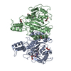
| ||||||||
|---|---|---|---|---|---|---|---|---|---|
| 1 | 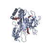
| ||||||||
| 2 | 
| ||||||||
| Unit cell |
|
-Components
| #1: Protein | Mass: 37918.414 Da / Num. of mol.: 2 / Source method: isolated from a natural source Source: (natural) Streptomyces mobaraensis NBRC 13819 = DSM 40847 (bacteria)References: UniProt: M3C567 #2: Protein/peptide | Mass: 1004.178 Da / Num. of mol.: 2 / Source method: obtained synthetically Details: chloroacetylated serine peptide derived from SPI aa1-10, Glutamin6 was replace by a chloroacetylated serine Source: (synth.) Streptomyces mobaraensis (bacteria) / References: UniProt: P86242*PLUS#3: Chemical | ChemComp-FLC / |   Mass: 189.100 Da / Num. of mol.: 1 / Source method: obtained synthetically / Formula: C6H5O7 Mass: 189.100 Da / Num. of mol.: 1 / Source method: obtained synthetically / Formula: C6H5O7#4: Chemical | ChemComp-PEG /   Mass: 106.120 Da / Num. of mol.: 5 / Source method: obtained synthetically / Formula: C4H10O3 Mass: 106.120 Da / Num. of mol.: 5 / Source method: obtained synthetically / Formula: C4H10O3#5: Water | ChemComp-HOH / |  Mass: 18.015 Da / Num. of mol.: 277 / Source method: isolated from a natural source / Formula: H2O Mass: 18.015 Da / Num. of mol.: 277 / Source method: isolated from a natural source / Formula: H2OHas protein modification | Y | |
|---|
-Experimental details
-Experiment
| Experiment | Method: X-RAY DIFFRACTION / Number of used crystals: 1 |
|---|
- Sample preparation
| Crystal | Density Matthews: 2.44 Å3/Da / Density % sol: 49.67 % |
|---|---|
| Crystal grow | Temperature: 292.15 K / Method: vapor diffusion, sitting drop Details: 38% v/v PEG400, 70 mM di-Sodium hydrogen phosphate, 100 mM Sodium phosphate citrate, pH 4.4 |
-Data collection
| Diffraction | Mean temperature: 100 K | ||||||||||||||||||||||||||||||
|---|---|---|---|---|---|---|---|---|---|---|---|---|---|---|---|---|---|---|---|---|---|---|---|---|---|---|---|---|---|---|---|
| Diffraction source | Source: SYNCHROTRON / Site: SLS  / Beamline: X06DA / Wavelength: 1 Å / Beamline: X06DA / Wavelength: 1 Å | ||||||||||||||||||||||||||||||
| Detector | Type: DECTRIS PILATUS 2M-F / Detector: PIXEL / Date: Jun 14, 2017 | ||||||||||||||||||||||||||||||
| Radiation | Protocol: SINGLE WAVELENGTH / Monochromatic (M) / Laue (L): M / Scattering type: x-ray | ||||||||||||||||||||||||||||||
| Radiation wavelength | Wavelength: 1 Å / Relative weight: 1 | ||||||||||||||||||||||||||||||
| Reflection | Resolution: 2.25→47 Å / Num. obs: 35089 / % possible obs: 98.9 % / Redundancy: 6.9 % / Biso Wilson estimate: 24.71 Å2 / CC1/2: 0.992 / Rmerge(I) obs: 0.196 / Rpim(I) all: 0.08 / Rrim(I) all: 0.212 / Net I/σ(I): 8.1 / Num. measured all: 241385 / Scaling rejects: 2 | ||||||||||||||||||||||||||||||
| Reflection shell | Diffraction-ID: 1
|
-Phasing
| Phasing | Method: molecular replacement | |||||||||
|---|---|---|---|---|---|---|---|---|---|---|
| Phasing MR |
|
- Processing
| Software |
| ||||||||||||||||||||||||||||||||||||||||||||||||||||||||||||||||||||||||||||||||||||||||||||||||||
|---|---|---|---|---|---|---|---|---|---|---|---|---|---|---|---|---|---|---|---|---|---|---|---|---|---|---|---|---|---|---|---|---|---|---|---|---|---|---|---|---|---|---|---|---|---|---|---|---|---|---|---|---|---|---|---|---|---|---|---|---|---|---|---|---|---|---|---|---|---|---|---|---|---|---|---|---|---|---|---|---|---|---|---|---|---|---|---|---|---|---|---|---|---|---|---|---|---|---|---|
| Refinement | Method to determine structure: MOLECULAR REPLACEMENT Starting model: 1iu4 Resolution: 2.25→47.001 Å / SU ML: 0.33 / Cross valid method: THROUGHOUT / σ(F): 1.35 / Phase error: 29.66 / Stereochemistry target values: ML
| ||||||||||||||||||||||||||||||||||||||||||||||||||||||||||||||||||||||||||||||||||||||||||||||||||
| Solvent computation | Shrinkage radii: 0.9 Å / VDW probe radii: 1.11 Å / Solvent model: FLAT BULK SOLVENT MODEL | ||||||||||||||||||||||||||||||||||||||||||||||||||||||||||||||||||||||||||||||||||||||||||||||||||
| Displacement parameters | Biso max: 74.05 Å2 / Biso mean: 25.9491 Å2 / Biso min: 11.43 Å2 | ||||||||||||||||||||||||||||||||||||||||||||||||||||||||||||||||||||||||||||||||||||||||||||||||||
| Refinement step | Cycle: final / Resolution: 2.25→47.001 Å
| ||||||||||||||||||||||||||||||||||||||||||||||||||||||||||||||||||||||||||||||||||||||||||||||||||
| LS refinement shell | Refine-ID: X-RAY DIFFRACTION / Rfactor Rfree error: 0 / Total num. of bins used: 13
|
