 Movie
Movie Controller


+ Open data

- Basic information
| Entry | Database: PDB / ID: 1iu4 | ||||||
|---|---|---|---|---|---|---|---|
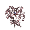
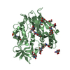
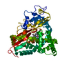
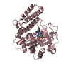
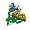
| Title | Crystal Structure Analysis of the Microbial Transglutaminase | ||||||
 Components Components | microbial transglutaminase | ||||||
 Keywords Keywords | TRANSFERASE / ALPHA-BETA | ||||||
| Function / homology |  Function and homology information Function and homology informationprotein-glutamine gamma-glutamyltransferase / protein-glutamine gamma-glutamyltransferase activity Similarity search - Function | ||||||
| Biological species |  Streptomyces mobaraensis (bacteria) Streptomyces mobaraensis (bacteria) | ||||||
| Method |  X-RAY DIFFRACTION / SYNCHROTRON / MIR / Resolution: 2.4 Å X-RAY DIFFRACTION / SYNCHROTRON / MIR / Resolution: 2.4 Å | ||||||
 Authors Authors | Kashiwagi, T. / Yokoyama, K. / Ishikawa, K. / Ono, K. / Ejima, D. / Matsui, H. / Suzuki, E. | ||||||
 Citation Citation | Journal: J.Biol.Chem. / Year: 2002 Title: Crystal structure of microbial transglutaminase from Streptoverticillium mobaraense Authors: Kashiwagi, T. / Yokoyama, K. / Ishikawa, K. / Ono, K. / Ejima, D. / Matsui, H. / Suzuki, E. | ||||||
| History |
|
- Structure visualization
| Structure viewer | Molecule: MolmilJmol/JSmol |
|---|
- Downloads & links
-Download
| PDBx/mmCIF format | 1iu4.cif.gz | 281.4 KB | Display | PDBx/mmCIF format |
|---|---|---|---|---|
| PDB format | pdb1iu4.ent.gz | 228.1 KB | Display | PDB format |
| PDBx/mmJSON format | 1iu4.json.gz | Tree view | PDBx/mmJSON format | |
| Others |  Other downloads Other downloads |
-Validation report
| Arichive directory | https://data.pdbj.org/pub/pdb/validation_reports/iu/1iu4ftp://data.pdbj.org/pub/pdb/validation_reports/iu/1iu4 | HTTPS FTP |
|---|
-Related structure data
| Similar structure data |
|---|


-Links
 PDBj
PDBj
- Assembly
| Deposited unit | 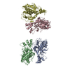
| ||||||||
|---|---|---|---|---|---|---|---|---|---|
| 1 | 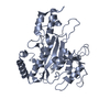
| ||||||||
| 2 | 
| ||||||||
| 3 | 
| ||||||||
| 4 | 
| ||||||||
| Unit cell |
| ||||||||
| Details | The protein acts as a monomer. |
-Components
| #1: Protein | Mass: 37918.414 Da / Num. of mol.: 4 / Fragment: residues 1-331 Source method: isolated from a genetically manipulated source Source: (gene. exp.) Streptomyces mobaraensis (bacteria) / Production host: References: UniProt: P81453, protein-glutamine gamma-glutamyltransferase #2: Water | ChemComp-HOH / |  Mass: 18.015 Da / Num. of mol.: 550 / Source method: isolated from a natural source / Formula: H2O Mass: 18.015 Da / Num. of mol.: 550 / Source method: isolated from a natural source / Formula: H2O |
|---|
-Experimental details
-Experiment
| Experiment | Method: X-RAY DIFFRACTION / Number of used crystals: 1 |
|---|
- Sample preparation
| Crystal | Density Matthews: 2.39 Å3/Da / Density % sol: 48.57 % | |||||||||||||||||||||||||||||||||||
|---|---|---|---|---|---|---|---|---|---|---|---|---|---|---|---|---|---|---|---|---|---|---|---|---|---|---|---|---|---|---|---|---|---|---|---|---|
| Crystal grow | Temperature: 293 K / Method: vapor diffusion, hanging drop / pH: 5 Details: PEG1000, calcium chloride, cacodylate, pH 5.0, VAPOR DIFFUSION, HANGING DROP, temperature 293K | |||||||||||||||||||||||||||||||||||
| Crystal grow | *PLUS Temperature: 20 ℃ | |||||||||||||||||||||||||||||||||||
| Components of the solutions | *PLUS
|
-Data collection
| Diffraction | Mean temperature: 100 K |
|---|---|
| Diffraction source | Source: SYNCHROTRON / Site: Photon Factory  / Beamline: BL-6B / Wavelength: 1 Å / Beamline: BL-6B / Wavelength: 1 Å |
| Detector | Type: FUJI / Detector: IMAGE PLATE / Date: Dec 2, 1999 |
| Radiation | Monochromator: Si(111) (Joham type) / Protocol: SINGLE WAVELENGTH / Monochromatic (M) / Laue (L): M / Scattering type: x-ray |
| Radiation wavelength | Wavelength: 1 Å / Relative weight: 1 |
| Reflection | Resolution: 2.4→40 Å / Num. all: 187008 / Num. obs: 55351 / % possible obs: 0.98 % / Observed criterion σ(F): 0 / Observed criterion σ(I): 0 / Redundancy: 3.38 % / Biso Wilson estimate: 26.25 Å2 / Rmerge(I) obs: 0.04 / Net I/σ(I): 12 |
| Reflection shell | Resolution: 2.4→2.44 Å / Rmerge(I) obs: 0.13 / % possible all: 0.921 |
| Reflection | *PLUS % possible obs: 0.98 % / Num. measured all: 187008 / Rmerge(I) obs: 0.04 |
| Reflection shell | *PLUS % possible obs: 92.1 % / Rmerge(I) obs: 0.13 |
- Processing
| Software |
| |||||||||||||||||||||||||
|---|---|---|---|---|---|---|---|---|---|---|---|---|---|---|---|---|---|---|---|---|---|---|---|---|---|---|
| Refinement | Method to determine structure: MIR / Resolution: 2.4→40 Å / σ(F): 2 Stereochemistry target values: standard set of QUANTA release98 (MSI)
| |||||||||||||||||||||||||
| Refinement step | Cycle: LAST / Resolution: 2.4→40 Å
| |||||||||||||||||||||||||
| Refine LS restraints |
| |||||||||||||||||||||||||
| Refinement | *PLUS % reflection Rfree: 5 % / Rfactor Rfree: 0.266 / Rfactor Rwork: 0.199 | |||||||||||||||||||||||||
| Solvent computation | *PLUS | |||||||||||||||||||||||||
| Displacement parameters | *PLUS |
