Monochromator: Si(111) / Protocol: SINGLE WAVELENGTH / Monochromatic (M) / Laue (L): M / Scattering type: x-ray
Radiation wavelength
Relative weight: 1
Reflection
Resolution: 2→33.273 Å / Num. all: 37270 / Num. obs: 27318 / % possible obs: 73.3 %
-
Processing
Software
Name
Version
Classification
Web-Ice
datacollection
CCP4
modelbuilding
REFMAC
5.6.0117
refinement
MOSFLM
datareduction
CCP4
phasing
Refinement
Method to determine structure: MOLECULAR REPLACEMENT / Resolution: 2→33.27 Å / Cor.coef. Fo:Fc: 0.953 / Cor.coef. Fo:Fc free: 0.912 / SU B: 3.82 / SU ML: 0.108 / Cross valid method: THROUGHOUT / σ(F): 2 / ESU R: 0.195 / ESU R Free: 0.172 / Stereochemistry target values: MAXIMUM LIKELIHOOD / Details: HYDROGENS HAVE BEEN USED IF PRESENT IN THE INPUT
Rfactor
Num. reflection
% reflection
Selection details
Rfree
0.22532
1353
5 %
RANDOM
Rwork
0.16992
-
-
-
all
0.17272
27318
-
-
obs
0.16992
25613
98.7 %
-
Solvent computation
Ion probe radii: 0.8 Å / Shrinkage radii: 0.8 Å / VDW probe radii: 1.2 Å / Solvent model: MASK
Displacement parameters
Biso mean: 19.52 Å2
Baniso -1
Baniso -2
Baniso -3
1-
0.02 Å2
0 Å2
0.06 Å2
2-
-
-0.03 Å2
-0 Å2
3-
-
-
0.04 Å2
Refinement step
Cycle: LAST / Resolution: 2→33.27 Å
Protein
Nucleic acid
Ligand
Solvent
Total
Num. atoms
3252
0
76
179
3507
Refine LS restraints
Refine-ID
Type
Dev ideal
Dev ideal target
Number
X-RAY DIFFRACTION
r_bond_refined_d
0.018
0.019
3496
X-RAY DIFFRACTION
r_angle_refined_deg
2.403
1.995
4788
X-RAY DIFFRACTION
r_dihedral_angle_1_deg
6.068
5
432
X-RAY DIFFRACTION
r_dihedral_angle_2_deg
35.347
24.121
165
X-RAY DIFFRACTION
r_dihedral_angle_3_deg
15.047
15
578
X-RAY DIFFRACTION
r_dihedral_angle_4_deg
12.659
15
26
X-RAY DIFFRACTION
r_chiral_restr
0.137
0.2
511
X-RAY DIFFRACTION
r_gen_planes_refined
0.012
0.021
2734
LS refinement shell
Resolution: 2→2.052 Å / Total num. of bins used: 20
Rfactor
Num. reflection
% reflection
Rfree
0.341
94
-
Rwork
0.216
1846
-
obs
-
-
98.93 %
+
About Yorodumi
-
News
-
Feb 9, 2022. New format data for meta-information of EMDB entries
New format data for meta-information of EMDB entries
Version 3 of the EMDB header file is now the official format.
The previous official version 1.9 will be removed from the archive.
In the structure databanks used in Yorodumi, some data are registered as the other names, "COVID-19 virus" and "2019-nCoV". Here are the details of the virus and the list of structure data.
Jan 31, 2019. EMDB accession codes are about to change! (news from PDBe EMDB page)
EMDB accession codes are about to change! (news from PDBe EMDB page)
The allocation of 4 digits for EMDB accession codes will soon come to an end. Whilst these codes will remain in use, new EMDB accession codes will include an additional digit and will expand incrementally as the available range of codes is exhausted. The current 4-digit format prefixed with “EMD-” (i.e. EMD-XXXX) will advance to a 5-digit format (i.e. EMD-XXXXX), and so on. It is currently estimated that the 4-digit codes will be depleted around Spring 2019, at which point the 5-digit format will come into force.
The EM Navigator/Yorodumi systems omit the EMD- prefix.
Related info.:Q: What is EMD? / ID/Accession-code notation in Yorodumi/EM Navigator
Yorodumi is a browser for structure data from EMDB, PDB, SASBDB, etc.
This page is also the successor to EM Navigator detail page, and also detail information page/front-end page for Omokage search.
The word "yorodu" (or yorozu) is an old Japanese word meaning "ten thousand". "mi" (miru) is to see.
Related info.:EMDB / PDB / SASBDB / Comparison of 3 databanks / Yorodumi Search / Aug 31, 2016. New EM Navigator & Yorodumi / Yorodumi Papers / Jmol/JSmol / Function and homology information / Changes in new EM Navigator and Yorodumi
 Movie
Movie Controller

 Yorodumi
 Open data
 Basic information
 Components
 Keywords
 Function and homology information
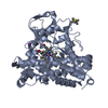
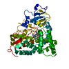
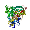
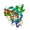
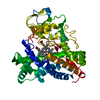
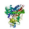
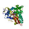
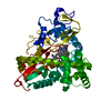
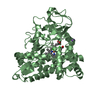
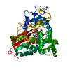
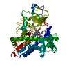
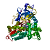
 Pseudomonas putida (bacteria)
 X-RAY DIFFRACTION /
  Authors
 Citation
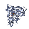
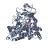
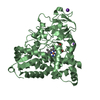
 Structure visualization
 Downloads & links
 Other downloads


 PDBj
PDBj
 Assembly
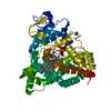


 Mass: 616.487 Da / Num. of mol.: 1 / Source method: obtained synthetically / Formula: C34H32FeN4O4
 Mass: 259.302 Da / Num. of mol.: 1 / Source method: obtained synthetically / Formula: C18H13NO
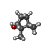
 Mass: 152.233 Da / Num. of mol.: 1 / Source method: obtained synthetically / Formula: C10H16O
 Mass: 39.098 Da / Num. of mol.: 2 / Source method: obtained synthetically / Formula: K
 Sample preparation
 / Beamline: BL7-1
 Processing