 Movie
Movie Controller

[English] 日本語
 Yorodumi
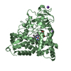
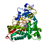
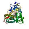
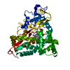
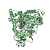
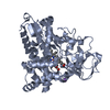
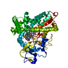
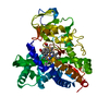
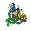
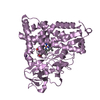
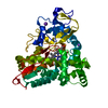
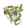
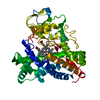
- PDB-2fe6: P450CAM from Pseudomonas putida reconstituted with manganic proto... -
+ Open data

- Basic information
| Entry | Database: PDB / ID: 2fe6 | ||||||
|---|---|---|---|---|---|---|---|
| Title | P450CAM from Pseudomonas putida reconstituted with manganic protoporphyrin IX | ||||||
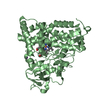
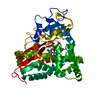
 Components Components | Cytochrome P450-cam | ||||||
 Keywords Keywords | OXIDOREDUCTASE / MONO-OXYGENASE / HEME / Manganic / Substrate-Free | ||||||
| Function / homology |  Function and homology information Function and homology informationcamphor 5-monooxygenase / camphor 5-monooxygenase activity / (+)-camphor catabolic process / iron ion binding / heme binding / cytoplasm Similarity search - Function | ||||||
| Biological species |  Pseudomonas putida (bacteria) Pseudomonas putida (bacteria) | ||||||
| Method |  X-RAY DIFFRACTION / SYNCHROTRON / FOURIER SYNTHESIS / Resolution: 1.5 Å X-RAY DIFFRACTION / SYNCHROTRON / FOURIER SYNTHESIS / Resolution: 1.5 Å | ||||||
 Authors Authors | von Koenig, K. / Makris, T.M. / Sligar, S.G. / Schlichting, I. | ||||||
 Citation Citation | Journal: J.Inorg.Biochem. / Year: 2006 Title: The status of high-valent metal oxo complexes in the P450 cytochromes. Authors: Makris, T.M. / Koenig, K. / Schlichting, I. / Sligar, S.G. | ||||||
| History |
|
- Structure visualization
| Structure viewer | Molecule: MolmilJmol/JSmol |
|---|
- Downloads & links
-Download
| PDBx/mmCIF format | 2fe6.cif.gz | 105.8 KB | Display | PDBx/mmCIF format |
|---|---|---|---|---|
| PDB format | pdb2fe6.ent.gz | 78.4 KB | Display | PDB format |
| PDBx/mmJSON format | 2fe6.json.gz | Tree view | PDBx/mmJSON format | |
| Others |  Other downloads Other downloads |
-Validation report
| Arichive directory | https://data.pdbj.org/pub/pdb/validation_reports/fe/2fe6ftp://data.pdbj.org/pub/pdb/validation_reports/fe/2fe6 | HTTPS FTP |
|---|
-Related structure data
| Related structure data |  2ferC  2feuC 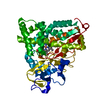 1yrdS S: Starting model for refinement C: citing same article ( |
|---|---|
| Similar structure data |


-Links
 PDBj
PDBj
- Assembly
| Deposited unit | 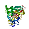
| ||||||||
|---|---|---|---|---|---|---|---|---|---|
| 1 |
| ||||||||
| Unit cell |
|
-Components
| #1: Protein | Mass: 47548.945 Da / Num. of mol.: 1 Source method: isolated from a genetically manipulated source Source: (gene. exp.) Pseudomonas putida (bacteria) / Gene: camC, cyp101 / Plasmid: pet28 / Production host: |
|---|---|
| #2: Chemical | ChemComp-K /   Mass: 39.098 Da / Num. of mol.: 1 / Source method: obtained synthetically / Formula: K Mass: 39.098 Da / Num. of mol.: 1 / Source method: obtained synthetically / Formula: K |
| #3: Chemical | ChemComp-MNR / 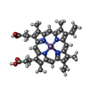  Mass: 615.580 Da / Num. of mol.: 1 / Source method: obtained synthetically / Formula: C34H32MnN4O4 Mass: 615.580 Da / Num. of mol.: 1 / Source method: obtained synthetically / Formula: C34H32MnN4O4 |
| #4: Water | ChemComp-HOH /  Mass: 18.015 Da / Num. of mol.: 430 / Source method: isolated from a natural source / Formula: H2O Mass: 18.015 Da / Num. of mol.: 430 / Source method: isolated from a natural source / Formula: H2O |
-Experimental details
-Experiment
| Experiment | Method: X-RAY DIFFRACTION / Number of used crystals: 1 |
|---|
- Sample preparation
| Crystal | Density Matthews: 2.87 Å3/Da / Density % sol: 57.11 % |
|---|---|
| Crystal grow | Temperature: 277 K / Method: vapor diffusion, sitting drop / pH: 7.4 Details: 7.5 MG/ML P450, 50 MM TRIS, 125 MM KCL, 13.5-15% PEG8000, 50 MM DTE, pH 7.4, VAPOR DIFFUSION, SITTING DROP, temperature 277K |
-Data collection
| Diffraction | Mean temperature: 90 K |
|---|---|
| Diffraction source | Source: SYNCHROTRON / Site: SLS  / Beamline: X10SA / Wavelength: 0.93 Å / Beamline: X10SA / Wavelength: 0.93 Å |
| Detector | Type: MARRESEARCH / Detector: CCD / Date: Jul 29, 2005 / Details: Si(111) monochromator, dynamically bendable mirror |
| Radiation | Monochromator: Si (111) / Protocol: SINGLE WAVELENGTH / Monochromatic (M) / Laue (L): M / Scattering type: x-ray |
| Radiation wavelength | Wavelength: 0.93 Å / Relative weight: 1 |
| Reflection | Resolution: 1.4→18 Å / Num. obs: 84598 / % possible obs: 96.6 % / Observed criterion σ(F): 0 / Observed criterion σ(I): 0 / Biso Wilson estimate: 19 Å2 / Rsym value: 0.086 / Net I/σ(I): 7.01 |
| Reflection shell | Resolution: 1.4→1.5 Å / Mean I/σ(I) obs: 2.38 / Num. unique all: 30433 / Num. unique obs: 18162 / Rsym value: 0.24 / % possible all: 93.4 |
- Processing
| Software |
| ||||||||||||||||||||||||||||
|---|---|---|---|---|---|---|---|---|---|---|---|---|---|---|---|---|---|---|---|---|---|---|---|---|---|---|---|---|---|
| Refinement | Method to determine structure: FOURIER SYNTHESIS Starting model: PDB ENTRY 1yrd Resolution: 1.5→18 Å / σ(F): 0 / Stereochemistry target values: Engh & Huber Details: initial parameters for Mn porphyrin IX from: Suslick, K.S. et al. (1991), Inorg. Chem. 30, 2311-17.
| ||||||||||||||||||||||||||||
| Solvent computation | Bsol: 52.904 Å2 | ||||||||||||||||||||||||||||
| Displacement parameters | Biso mean: 16.327 Å2
| ||||||||||||||||||||||||||||
| Refinement step | Cycle: LAST / Resolution: 1.5→18 Å
| ||||||||||||||||||||||||||||
| Refine LS restraints |
| ||||||||||||||||||||||||||||
| LS refinement shell | Resolution: 1.5→1.6 Å / Num. reflection obs: 26508 | ||||||||||||||||||||||||||||
| Xplor file |
|
