 Movie
Movie Controller


+ Open data

- Basic information
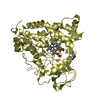
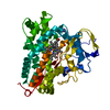
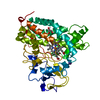
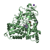
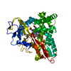
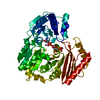
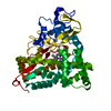
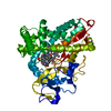
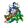
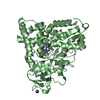
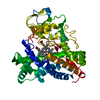
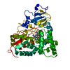
| Entry | Database: PDB / ID: 1noo | ||||||
|---|---|---|---|---|---|---|---|
| Title | CYTOCHROME P450-CAM COMPLEXED WITH 5-EXO-HYDROXYCAMPHOR | ||||||
 Components Components | CYTOCHROME P450-CAM | ||||||
 Keywords Keywords | OXIDOREDUCTASE (OXYGENASE) / MONOOXYGENASE | ||||||
| Function / homology |  Function and homology information Function and homology informationcamphor 5-monooxygenase / camphor 5-monooxygenase activity / (+)-camphor catabolic process / iron ion binding / heme binding / cytoplasm Similarity search - Function | ||||||
| Biological species |  Pseudomonas putida (bacteria) Pseudomonas putida (bacteria) | ||||||
| Method |  X-RAY DIFFRACTION / Resolution: 2.2 Å X-RAY DIFFRACTION / Resolution: 2.2 Å | ||||||
 Authors Authors | Li, H.Y. / Poulos, T.L. | ||||||
 Citation Citation | Journal: J.Am.Chem.Soc. / Year: 1995 Title: Crystal Structure of Cytochrome P450-Cam Complexed with its Catalytic Product, 5-Exo-Hydroxycamphor Authors: Li, H. / Narasimhulu, S. / Havran, L.M. / Winkler, J.D. / Poulos, T.L. #1: Journal: J.Mol.Biol. / Year: 1987Title: High-Resolution Crystal Structure of Cytochrome P450-Cam Authors: Poulos, T.L. / Finzel, B.C. / Howard, A.J. | ||||||
| History |
|
- Structure visualization
| Structure viewer | Molecule: MolmilJmol/JSmol |
|---|
- Downloads & links
-Download
| PDBx/mmCIF format | 1noo.cif.gz | 97.4 KB | Display | PDBx/mmCIF format |
|---|---|---|---|---|
| PDB format | pdb1noo.ent.gz | 74.1 KB | Display | PDB format |
| PDBx/mmJSON format | 1noo.json.gz | Tree view | PDBx/mmJSON format | |
| Others |  Other downloads Other downloads |
-Validation report
| Arichive directory | https://data.pdbj.org/pub/pdb/validation_reports/no/1nooftp://data.pdbj.org/pub/pdb/validation_reports/no/1noo | HTTPS FTP |
|---|
-Related structure data
| Similar structure data |
|---|


-Links
 PDBj
PDBj
- Assembly
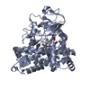
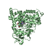
| Deposited unit | 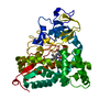
| ||||||||
|---|---|---|---|---|---|---|---|---|---|
| 1 |
| ||||||||
| Unit cell |
| ||||||||
| Atom site foot note | 1: CIS PROLINE - PRO 89 / 2: CIS PROLINE - PRO 100 / 3: CIS PROLINE - PRO 106 |
-Components
| #1: Protein | Mass: 46588.879 Da / Num. of mol.: 1 Source method: isolated from a genetically manipulated source Source: (gene. exp.) Pseudomonas putida (bacteria) / Production host: |
|---|---|
| #2: Chemical | ChemComp-HEM /   Mass: 616.487 Da / Num. of mol.: 1 / Source method: obtained synthetically / Formula: C34H32FeN4O4 Mass: 616.487 Da / Num. of mol.: 1 / Source method: obtained synthetically / Formula: C34H32FeN4O4 |
| #3: Chemical | ChemComp-CAH /   Mass: 168.233 Da / Num. of mol.: 1 / Source method: obtained synthetically / Formula: C10H16O2 Mass: 168.233 Da / Num. of mol.: 1 / Source method: obtained synthetically / Formula: C10H16O2 |
| #4: Water | ChemComp-HOH /  Mass: 18.015 Da / Num. of mol.: 214 / Source method: isolated from a natural source / Formula: H2O Mass: 18.015 Da / Num. of mol.: 214 / Source method: isolated from a natural source / Formula: H2O |
-Experimental details
-Experiment
| Experiment | Method: X-RAY DIFFRACTION / Number of used crystals: 1 |
|---|
- Sample preparation
| Crystal | Density Matthews: 2.22 Å3/Da / Density % sol: 44.57 % |
|---|---|
| Crystal grow | *PLUS Temperature: 7 ℃ / Method: free interface diffusion method |
| Components of the solutions | *PLUS Conc.: 50 mM / Common name: dithiothreitol |
-Data collection
| Diffraction | Mean temperature: 295 K |
|---|---|
| Diffraction source | Wavelength: 1.5418 Å |
| Detector | Type: SIEMENS-NICOLET X100 / Detector: AREA DETECTOR / Date: Oct 25, 1994 |
| Radiation | Monochromatic (M) / Laue (L): M / Scattering type: x-ray |
| Radiation wavelength | Wavelength: 1.5418 Å / Relative weight: 1 |
| Reflection | Resolution: 2.2→50 Å / Num. obs: 20570 / % possible obs: 94 % / Redundancy: 4 % / Rmerge(I) obs: 0.0975 |
| Reflection | *PLUS Rmerge(I) obs: 0.0975 |
- Processing
| Software |
| ||||||||||||||||||||||||||||||||||||||||||||||||||||||||||||
|---|---|---|---|---|---|---|---|---|---|---|---|---|---|---|---|---|---|---|---|---|---|---|---|---|---|---|---|---|---|---|---|---|---|---|---|---|---|---|---|---|---|---|---|---|---|---|---|---|---|---|---|---|---|---|---|---|---|---|---|---|---|
| Refinement | Resolution: 2.2→10 Å / σ(F): 2
| ||||||||||||||||||||||||||||||||||||||||||||||||||||||||||||
| Refine analyze | Luzzati coordinate error obs: 0.2 Å | ||||||||||||||||||||||||||||||||||||||||||||||||||||||||||||
| Refinement step | Cycle: LAST / Resolution: 2.2→10 Å
| ||||||||||||||||||||||||||||||||||||||||||||||||||||||||||||
| Refine LS restraints |
| ||||||||||||||||||||||||||||||||||||||||||||||||||||||||||||
| Software | *PLUS Name: X-PLOR / Classification: refinement | ||||||||||||||||||||||||||||||||||||||||||||||||||||||||||||
| Refinement | *PLUS | ||||||||||||||||||||||||||||||||||||||||||||||||||||||||||||
| Solvent computation | *PLUS | ||||||||||||||||||||||||||||||||||||||||||||||||||||||||||||
| Displacement parameters | *PLUS | ||||||||||||||||||||||||||||||||||||||||||||||||||||||||||||
| Refine LS restraints | *PLUS
|
