 Movie
Movie Controller


+ Open data

- Basic information
| Entry | Database: PDB / ID: 1p5g | ||||||
|---|---|---|---|---|---|---|---|
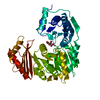
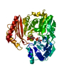
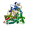
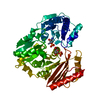
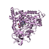
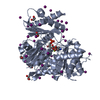
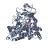
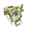
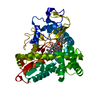
| Title | Enzyme-ligand complex of P. aeruginosa PMM/PGM | ||||||
 Components Components | Phosphomannomutase | ||||||
 Keywords Keywords | ISOMERASE / alpha/beta protein / phosphohexomutase / phosphoserine / enzyme-ligand complex / enzyme-metal complex | ||||||
| Function / homology |  Function and homology information Function and homology informationphosphomannomutase / phosphoglucomutase (alpha-D-glucose-1,6-bisphosphate-dependent) / phosphoglucomutase activity / phosphomannomutase activity / alginic acid biosynthetic process / O antigen biosynthetic process / GDP-mannose biosynthetic process / lipopolysaccharide core region biosynthetic process / magnesium ion binding Similarity search - Function | ||||||
| Biological species |   Pseudomonas aeruginosa (bacteria) Pseudomonas aeruginosa (bacteria) | ||||||
| Method |  X-RAY DIFFRACTION / SYNCHROTRON / MOLECULAR REPLACEMENT / Resolution: 1.61 Å X-RAY DIFFRACTION / SYNCHROTRON / MOLECULAR REPLACEMENT / Resolution: 1.61 Å | ||||||
 Authors Authors | Regni, C. / Tipton, P.A. / Beamer, L.J. | ||||||
 Citation Citation | Journal: Structure / Year: 2004 Title: Structural basis of diverse substrate recognition by the enzyme PMM/PGM from P. aeruginosa. Authors: Regni, C. / Naught, L. / Tipton, P.A. / Beamer, L.J. | ||||||
| History |
|
- Structure visualization
| Structure viewer | Molecule: MolmilJmol/JSmol |
|---|
- Downloads & links
-Download
| PDBx/mmCIF format | 1p5g.cif.gz | 112.9 KB | Display | PDBx/mmCIF format |
|---|---|---|---|---|
| PDB format | pdb1p5g.ent.gz | 84.8 KB | Display | PDB format |
| PDBx/mmJSON format | 1p5g.json.gz | Tree view | PDBx/mmJSON format | |
| Others |  Other downloads Other downloads |
-Validation report
| Arichive directory | https://data.pdbj.org/pub/pdb/validation_reports/p5/1p5gftp://data.pdbj.org/pub/pdb/validation_reports/p5/1p5g | HTTPS FTP |
|---|
-Related structure data
| Related structure data | 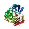 1p5dC 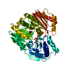 1pcjC  1pcmC 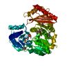 1k35S S: Starting model for refinement C: citing same article ( |
|---|---|
| Similar structure data |


-Links
 PDBj
PDBj- Assembly
| Deposited unit | 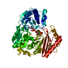
| ||||||||
|---|---|---|---|---|---|---|---|---|---|
| 1 |
| ||||||||
| Unit cell |
|
-Components
| #1: Protein | Mass: 50430.328 Da / Num. of mol.: 1 Source method: isolated from a genetically manipulated source Source: (gene. exp.) Pseudomonas aeruginosa (bacteria) / Gene: ALGC OR PA5322 / Plasmid: PET3-A / Production host: |
|---|---|
| #2: Sugar | ChemComp-G6P /   Type: D-saccharide, alpha linking / Mass: 260.136 Da / Num. of mol.: 1 Type: D-saccharide, alpha linking / Mass: 260.136 Da / Num. of mol.: 1Source method: isolated from a genetically manipulated source Formula: C6H13O9P |
| #3: Chemical | ChemComp-ZN /   Mass: 65.409 Da / Num. of mol.: 1 / Source method: obtained synthetically / Formula: Zn Mass: 65.409 Da / Num. of mol.: 1 / Source method: obtained synthetically / Formula: Zn |
| #4: Water | ChemComp-HOH /  Mass: 18.015 Da / Num. of mol.: 437 / Source method: isolated from a natural source / Formula: H2O Mass: 18.015 Da / Num. of mol.: 437 / Source method: isolated from a natural source / Formula: H2O |
| Has protein modification | Y |
-Experimental details
-Experiment
| Experiment | Method: X-RAY DIFFRACTION / Number of used crystals: 1 |
|---|
- Sample preparation
| Crystal | Density Matthews: 2.24 Å3/Da / Density % sol: 45.01 % | ||||||||||||||||||||||||||||||
|---|---|---|---|---|---|---|---|---|---|---|---|---|---|---|---|---|---|---|---|---|---|---|---|---|---|---|---|---|---|---|---|
| Crystal grow | Temperature: 293 K / Method: vapor diffusion, hanging drop / pH: 7.5 Details: Na K tartrate, Hepes, pH 7.5, VAPOR DIFFUSION, HANGING DROP, temperature 293K | ||||||||||||||||||||||||||||||
| Crystal grow | *PLUS pH: 7 / Method: vapor diffusion, hanging drop / Details: Regni, C.A., (2000) Acta Crystallogr, D56, 761. | ||||||||||||||||||||||||||||||
| Components of the solutions | *PLUS
|
-Data collection
| Diffraction | Mean temperature: 100 K |
|---|---|
| Diffraction source | Source: SYNCHROTRON / Site: APS  / Beamline: 14-BM-C / Wavelength: 0.9 Å / Beamline: 14-BM-C / Wavelength: 0.9 Å |
| Detector | Type: ADSC QUANTUM 4 / Detector: CCD / Date: Feb 8, 2003 / Details: mirrors |
| Radiation | Monochromator: Bent Ge(111) / Protocol: SINGLE WAVELENGTH / Monochromatic (M) / Laue (L): M / Scattering type: x-ray |
| Radiation wavelength | Wavelength: 0.9 Å / Relative weight: 1 |
| Reflection | Resolution: 1.6→25 Å / Num. all: 60200 / Num. obs: 59418 / % possible obs: 98.7 % / Observed criterion σ(F): 0 / Observed criterion σ(I): 0 / Redundancy: 12.9 % / Biso Wilson estimate: 17.4 Å2 / Rmerge(I) obs: 0.062 / Net I/σ(I): 41.8 |
| Reflection shell | Resolution: 1.6→1.66 Å / Rmerge(I) obs: 0.477 / Mean I/σ(I) obs: 5.6 / % possible all: 87.7 |
| Reflection shell | *PLUS % possible obs: 87.7 % |
- Processing
| Software |
| ||||||||||||||||||||||||||||||||||||||||||||||||||||||||||||||||||||||||||||||||||||||||||||||||||||
|---|---|---|---|---|---|---|---|---|---|---|---|---|---|---|---|---|---|---|---|---|---|---|---|---|---|---|---|---|---|---|---|---|---|---|---|---|---|---|---|---|---|---|---|---|---|---|---|---|---|---|---|---|---|---|---|---|---|---|---|---|---|---|---|---|---|---|---|---|---|---|---|---|---|---|---|---|---|---|---|---|---|---|---|---|---|---|---|---|---|---|---|---|---|---|---|---|---|---|---|---|---|
| Refinement | Method to determine structure: MOLECULAR REPLACEMENT Starting model: 1K35 Resolution: 1.61→25 Å / Cor.coef. Fo:Fc: 0.968 / Cor.coef. Fo:Fc free: 0.955 / SU B: 1.464 / SU ML: 0.052 / TLS residual ADP flag: LIKELY RESIDUAL / Cross valid method: THROUGHOUT / σ(F): 0 / σ(I): 0 / ESU R: 0.082 / ESU R Free: 0.081 / Stereochemistry target values: MAXIMUM LIKELIHOOD / Details: HYDROGENS HAVE BEEN ADDED IN THE RIDING POSITIONS
| ||||||||||||||||||||||||||||||||||||||||||||||||||||||||||||||||||||||||||||||||||||||||||||||||||||
| Solvent computation | Ion probe radii: 0.8 Å / Shrinkage radii: 0.8 Å / VDW probe radii: 1.4 Å / Solvent model: BABINET MODEL WITH MASK | ||||||||||||||||||||||||||||||||||||||||||||||||||||||||||||||||||||||||||||||||||||||||||||||||||||
| Displacement parameters | Biso mean: 14.015 Å2
| ||||||||||||||||||||||||||||||||||||||||||||||||||||||||||||||||||||||||||||||||||||||||||||||||||||
| Refinement step | Cycle: LAST / Resolution: 1.61→25 Å
| ||||||||||||||||||||||||||||||||||||||||||||||||||||||||||||||||||||||||||||||||||||||||||||||||||||
| Refine LS restraints |
| ||||||||||||||||||||||||||||||||||||||||||||||||||||||||||||||||||||||||||||||||||||||||||||||||||||
| LS refinement shell | Resolution: 1.61→1.695 Å / Rfactor Rfree error: 0.082 / Total num. of bins used: 10
| ||||||||||||||||||||||||||||||||||||||||||||||||||||||||||||||||||||||||||||||||||||||||||||||||||||
| Refinement TLS params. | Method: refined / Origin x: -49.7804 Å / Origin y: -18.1648 Å / Origin z: 10.9024 Å
| ||||||||||||||||||||||||||||||||||||||||||||||||||||||||||||||||||||||||||||||||||||||||||||||||||||
| Refinement | *PLUS Highest resolution: 1.6 Å / Lowest resolution: 25 Å / Rfactor Rfree: 0.19 / Rfactor Rwork: 0.16 | ||||||||||||||||||||||||||||||||||||||||||||||||||||||||||||||||||||||||||||||||||||||||||||||||||||
| Solvent computation | *PLUS | ||||||||||||||||||||||||||||||||||||||||||||||||||||||||||||||||||||||||||||||||||||||||||||||||||||
| Displacement parameters | *PLUS | ||||||||||||||||||||||||||||||||||||||||||||||||||||||||||||||||||||||||||||||||||||||||||||||||||||
| Refine LS restraints | *PLUS
|
