| 登録情報 | データベース: PDB / ID: 2h4l
|
|---|
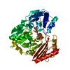
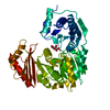
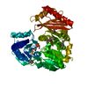
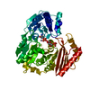
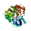
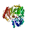
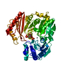
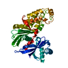
| タイトル | Complex of PMM/PGM with ribose 1-phosphate |
|---|
 要素 要素 | Phosphomannomutase/phosphoglucomutase |
|---|
 キーワード キーワード | ISOMERASE / PROTEIN-LIGAND COMPLEX / SLOW SUBSTRATE / RIBOSE 1-PHOSPHATE |
|---|
| 機能・相同性 |  機能・相同性情報 機能・相同性情報
phosphomannomutase / phosphoglucomutase (alpha-D-glucose-1,6-bisphosphate-dependent) / phosphoglucomutase activity / phosphomannomutase activity / alginic acid biosynthetic process / O antigen biosynthetic process / GDP-mannose biosynthetic process / lipopolysaccharide core region biosynthetic process / magnesium ion binding類似検索 - 分子機能 Alpha-D-phosphohexomutase, C-terminal / Phosphoglucomutase/phosphomannomutase, C-terminal domain / Alpha-D-Glucose-1,6-Bisphosphate; Chain A, domain 3 / Alpha-D-Glucose-1,6-Bisphosphate, subunit A, domain 3 / Alpha-D-phosphohexomutase, C-terminal domain / Alpha-D-phosphohexomutase superfamily / Alpha-D-phosphohexomutase, conserved site / Phosphoglucomutase and phosphomannomutase phosphoserine signature. / Alpha-D-phosphohexomutase, alpha/beta/alpha domain II / Alpha-D-phosphohexomutase, alpha/beta/alpha domain III ...Alpha-D-phosphohexomutase, C-terminal / Phosphoglucomutase/phosphomannomutase, C-terminal domain / Alpha-D-Glucose-1,6-Bisphosphate; Chain A, domain 3 / Alpha-D-Glucose-1,6-Bisphosphate, subunit A, domain 3 / Alpha-D-phosphohexomutase, C-terminal domain / Alpha-D-phosphohexomutase superfamily / Alpha-D-phosphohexomutase, conserved site / Phosphoglucomutase and phosphomannomutase phosphoserine signature. / Alpha-D-phosphohexomutase, alpha/beta/alpha domain II / Alpha-D-phosphohexomutase, alpha/beta/alpha domain III / Phosphoglucomutase/phosphomannomutase, alpha/beta/alpha domain II / Phosphoglucomutase/phosphomannomutase, alpha/beta/alpha domain III / Alpha-D-phosphohexomutase, alpha/beta/alpha domain I / Alpha-D-phosphohexomutase, alpha/beta/alpha I/II/III / Alpha-D-phosphohexomutase, C-terminal domain superfamily / Phosphoglucomutase/phosphomannomutase, alpha/beta/alpha domain I / TATA-Binding Protein / 2-Layer Sandwich / 3-Layer(aba) Sandwich / Alpha Beta類似検索 - ドメイン・相同性 1-O-phosphono-alpha-D-ribofuranose / Phosphomannomutase/phosphoglucomutase類似検索 - 構成要素 |
|---|
| 生物種 |   Pseudomonas aeruginosa (緑膿菌) Pseudomonas aeruginosa (緑膿菌) |
|---|
| 手法 |  X線回折 / シンクロトロン / フーリエ合成 / 解像度: 2.4 Å X線回折 / シンクロトロン / フーリエ合成 / 解像度: 2.4 Å |
|---|
 データ登録者 データ登録者 | Beamer, L.J. |
|---|
 引用 引用 | ジャーナル: Acta Crystallogr.,Sect.F / 年: 2006
タイトル: Complexes of the enzyme phosphomannomutase/phosphoglucomutase with a slow substrate and an inhibitor.
著者: Regni, C. / Shackelford, G.S. / Beamer, L.J. |
|---|
| 履歴 | | 登録 | 2006年5月24日 | 登録サイト: RCSB / 処理サイト: RCSB |
|---|
| 改定 1.0 | 2006年8月8日 | Provider: repository / タイプ: Initial release |
|---|
| 改定 1.1 | 2008年5月1日 | Group: Version format compliance |
|---|
| 改定 1.2 | 2011年7月13日 | Group: Advisory / Version format compliance |
|---|
| 改定 2.0 | 2020年7月29日 | Group: Atomic model / Data collection ...Atomic model / Data collection / Database references / Derived calculations / Structure summary
カテゴリ: atom_site / chem_comp ...atom_site / chem_comp / entity / pdbx_chem_comp_identifier / pdbx_entity_nonpoly / pdbx_struct_conn_angle / struct_conn / struct_ref_seq_dif / struct_site / struct_site_gen
Item: _atom_site.auth_atom_id / _atom_site.label_atom_id ..._atom_site.auth_atom_id / _atom_site.label_atom_id / _chem_comp.mon_nstd_flag / _chem_comp.name / _chem_comp.type / _entity.pdbx_description / _pdbx_entity_nonpoly.name / _pdbx_struct_conn_angle.ptnr1_auth_comp_id / _pdbx_struct_conn_angle.ptnr1_auth_seq_id / _pdbx_struct_conn_angle.ptnr1_label_atom_id / _pdbx_struct_conn_angle.ptnr1_label_comp_id / _pdbx_struct_conn_angle.ptnr1_label_seq_id / _pdbx_struct_conn_angle.ptnr3_auth_comp_id / _pdbx_struct_conn_angle.ptnr3_auth_seq_id / _pdbx_struct_conn_angle.ptnr3_label_atom_id / _pdbx_struct_conn_angle.ptnr3_label_comp_id / _pdbx_struct_conn_angle.ptnr3_label_seq_id / _pdbx_struct_conn_angle.value / _struct_conn.pdbx_dist_value / _struct_conn.pdbx_leaving_atom_flag / _struct_conn.ptnr1_auth_comp_id / _struct_conn.ptnr1_auth_seq_id / _struct_conn.ptnr1_label_asym_id / _struct_conn.ptnr1_label_atom_id / _struct_conn.ptnr1_label_comp_id / _struct_conn.ptnr1_label_seq_id / _struct_conn.ptnr2_auth_comp_id / _struct_conn.ptnr2_auth_seq_id / _struct_conn.ptnr2_label_asym_id / _struct_conn.ptnr2_label_atom_id / _struct_conn.ptnr2_label_comp_id / _struct_conn.ptnr2_label_seq_id / _struct_ref_seq_dif.details
解説: Carbohydrate remediation / Provider: repository / タイプ: Remediation |
|---|
| 改定 2.1 | 2023年8月30日 | Group: Data collection / Database references ...Data collection / Database references / Refinement description / Structure summary
カテゴリ: chem_comp / chem_comp_atom ...chem_comp / chem_comp_atom / chem_comp_bond / database_2 / pdbx_initial_refinement_model
Item: _chem_comp.pdbx_synonyms / _database_2.pdbx_DOI / _database_2.pdbx_database_accession |
|---|
| 改定 2.2 | 2024年10月9日 | Group: Structure summary
カテゴリ: pdbx_entry_details / pdbx_modification_feature |
|---|
|
|---|
 ムービー
ムービー コントローラー


 データを開く
 基本情報
基本情報 構造の表示
構造の表示 ダウンロードとリンク
 その他のダウンロード
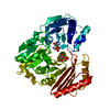


 PDBj
PDBj 集合体
集合体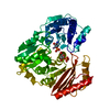
 タイプ: D-saccharide, alpha linking / 分子量: 230.110 Da / 分子数: 1 / 由来タイプ: 組換発現 / 式: C5H11O8P
 分子量: 65.409 Da / 分子数: 1 / 由来タイプ: 合成 / 式: Zn
 分子量: 18.015 Da / 分子数: 144 / 由来タイプ: 天然 / 式: H2O
 試料調製
試料調製 / ビームライン: 14-BM-C / 波長: 1.0398 Å
 解析
解析