- PDB-2fkm: PMM/PGM S108D mutant with alpha-d-glucose 1,6-bisphosphate bound -
+
Open data
ID or keywords:
Loading...
-
Basic information
Entry
Database: PDB / ID: 2fkm
Title
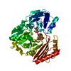
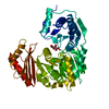
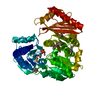
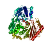
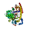
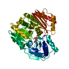
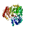
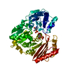
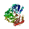
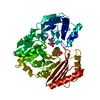
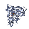
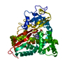
PMM/PGM S108D mutant with alpha-d-glucose 1,6-bisphosphate bound
Components
Phosphomannomutase/phosphoglucomutase
Keywords
ISOMERASE / alpha/beta protein / enzyme-metal complex / enzyme-ligand complex
Function / homology
Function and homology information
phosphomannomutase / phosphoglucomutase (alpha-D-glucose-1,6-bisphosphate-dependent) / phosphoglucomutase activity / phosphomannomutase activity / alginic acid biosynthetic process / O antigen biosynthetic process / GDP-mannose biosynthetic process / lipopolysaccharide core region biosynthetic process / magnesium ion binding Similarity search - Function
Alpha-D-phosphohexomutase, C-terminal / Phosphoglucomutase/phosphomannomutase, C-terminal domain / Alpha-D-Glucose-1,6-Bisphosphate; Chain A, domain 3 / Alpha-D-Glucose-1,6-Bisphosphate, subunit A, domain 3 / Alpha-D-phosphohexomutase, C-terminal domain / Alpha-D-phosphohexomutase superfamily / Alpha-D-phosphohexomutase, conserved site / Phosphoglucomutase and phosphomannomutase phosphoserine signature. / Alpha-D-phosphohexomutase, alpha/beta/alpha domain II / Alpha-D-phosphohexomutase, alpha/beta/alpha domain III ...Alpha-D-phosphohexomutase, C-terminal / Phosphoglucomutase/phosphomannomutase, C-terminal domain / Alpha-D-Glucose-1,6-Bisphosphate; Chain A, domain 3 / Alpha-D-Glucose-1,6-Bisphosphate, subunit A, domain 3 / Alpha-D-phosphohexomutase, C-terminal domain / Alpha-D-phosphohexomutase superfamily / Alpha-D-phosphohexomutase, conserved site / Phosphoglucomutase and phosphomannomutase phosphoserine signature. / Alpha-D-phosphohexomutase, alpha/beta/alpha domain II / Alpha-D-phosphohexomutase, alpha/beta/alpha domain III / Phosphoglucomutase/phosphomannomutase, alpha/beta/alpha domain II / Phosphoglucomutase/phosphomannomutase, alpha/beta/alpha domain III / Alpha-D-phosphohexomutase, alpha/beta/alpha domain I / Alpha-D-phosphohexomutase, alpha/beta/alpha I/II/III / Alpha-D-phosphohexomutase, C-terminal domain superfamily / Phosphoglucomutase/phosphomannomutase, alpha/beta/alpha domain I / TATA-Binding Protein / 2-Layer Sandwich / 3-Layer(aba) Sandwich / Alpha Beta Similarity search - Domain/homology
Mass: 18.015 Da / Num. of mol.: 234 / Source method: isolated from a natural source / Formula: H2O
-
Experimental details
-
Experiment
Experiment
Method: X-RAY DIFFRACTION / Number of used crystals: 1
-
Sample preparation
Crystal
Density Matthews: 2.07 Å3/Da / Density % sol: 40.68 %
Crystal grow
Temperature: 298 K / Method: vapor diffusion, hanging drop / pH: 7.5 Details: 50-60% sat Na K tartrate, .1 M Na Hepes pH 7.5 seeding used, VAPOR DIFFUSION, HANGING DROP, temperature 298K
-
Data collection
Diffraction
Mean temperature: 100 K
Diffraction source
Source: SYNCHROTRON / Site: ALS / Beamline: 4.2.2 / Wavelength: 0.97909 Å
Detector
Type: NOIR-1 / Detector: CCD / Date: Mar 4, 2005
Radiation
Monochromator: double crystal / Protocol: SINGLE WAVELENGTH / Monochromatic (M) / Laue (L): M / Scattering type: x-ray
In the structure databanks used in Yorodumi, some data are registered as the other names, "COVID-19 virus" and "2019-nCoV". Here are the details of the virus and the list of structure data.
Jan 31, 2019. EMDB accession codes are about to change! (news from PDBe EMDB page)
EMDB accession codes are about to change! (news from PDBe EMDB page)
The allocation of 4 digits for EMDB accession codes will soon come to an end. Whilst these codes will remain in use, new EMDB accession codes will include an additional digit and will expand incrementally as the available range of codes is exhausted. The current 4-digit format prefixed with “EMD-” (i.e. EMD-XXXX) will advance to a 5-digit format (i.e. EMD-XXXXX), and so on. It is currently estimated that the 4-digit codes will be depleted around Spring 2019, at which point the 5-digit format will come into force.
The EM Navigator/Yorodumi systems omit the EMD- prefix.
Related info.:Q: What is EMD? / ID/Accession-code notation in Yorodumi/EM Navigator
Yorodumi is a browser for structure data from EMDB, PDB, SASBDB, etc.
This page is also the successor to EM Navigator detail page, and also detail information page/front-end page for Omokage search.
The word "yorodu" (or yorozu) is an old Japanese word meaning "ten thousand". "mi" (miru) is to see.
Related info.:EMDB / PDB / SASBDB / Comparison of 3 databanks / Yorodumi Search / Aug 31, 2016. New EM Navigator & Yorodumi / Yorodumi Papers / Jmol/JSmol / Function and homology information / Changes in new EM Navigator and Yorodumi
 Movie
Movie Controller


 Open data
 Basic information
 Components
 Keywords
 Function and homology information
 Pseudomonas aeruginosa (bacteria)
 X-RAY DIFFRACTION /
  Authors
 Citation
 Structure visualization
 Downloads & links
 Other downloads
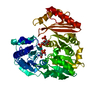


 PDBj
PDBj Assembly
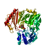
 Type: D-saccharide / Mass: 339.108 Da / Num. of mol.: 1
 Mass: 65.409 Da / Num. of mol.: 1 / Source method: obtained synthetically / Formula: Zn
 Mass: 18.015 Da / Num. of mol.: 234 / Source method: isolated from a natural source / Formula: H2O
 Sample preparation
 / Beamline: 4.2.2 / Wavelength: 0.97909 Å
 Processing