 Movie
Movie Controller


+ Open data

- Basic information
| Entry | Database: PDB / ID: 1uyu | ||||||
|---|---|---|---|---|---|---|---|
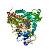
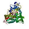
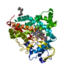
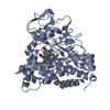
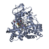
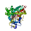
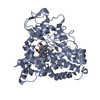
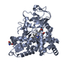
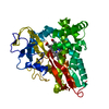
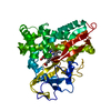
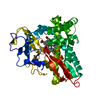
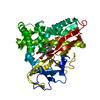
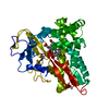
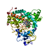
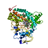
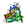
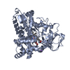
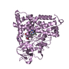
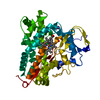
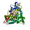
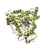
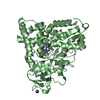
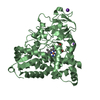
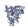
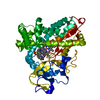
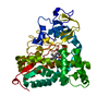
| Title | Xenon COMPLEX OF wildtype P450CAM FROM PSEUDOMONAS PUTIDA | ||||||
 Components Components | CYTOCHROME P450-CAM | ||||||
 Keywords Keywords | OXIDOREDUCTASE / MONO-OXYGENASE / HEME / ELECTRON TRANSPORT | ||||||
| Function / homology |  Function and homology information Function and homology informationcamphor 5-monooxygenase / camphor 5-monooxygenase activity / (+)-camphor catabolic process / iron ion binding / heme binding / cytoplasm Similarity search - Function | ||||||
| Biological species |  PSEUDOMONAS PUTIDA (bacteria) PSEUDOMONAS PUTIDA (bacteria) | ||||||
| Method |  X-RAY DIFFRACTION / SYNCHROTRON / MOLECULAR REPLACEMENT / Resolution: 2 Å X-RAY DIFFRACTION / SYNCHROTRON / MOLECULAR REPLACEMENT / Resolution: 2 Å | ||||||
 Authors Authors | Wade, R.C. / Winn, P.J. / Schlichting, I. / Sudarko, X. | ||||||
 Citation Citation | Journal: J.Inorg.Biochem. / Year: 2004 Title: A Survey of Active Site Access Channels in Cytochromes P450 Authors: Wade, R.C. / Winn, P.J. / Schlichting, I. #1: Journal: Annu.Rev.Biochem. / Year: 2004 Title: Structural Aspects of Ligand Binding to and Electron Transfer in Bacterial and Fungal P450S Authors: Pylypenko, O. / Schlichting, I. #2: Journal: J.Mol.Biol. / Year: 1987Title: High-Resolution Crystal Structure of Cytochrome P450Cam Authors: Poulos, T.L. / Finzel, B.C. / Howard, A.J. | ||||||
| History |
|
- Structure visualization
| Structure viewer | Molecule: MolmilJmol/JSmol |
|---|
- Downloads & links
-Download
| PDBx/mmCIF format | 1uyu.cif.gz | 186.4 KB | Display | PDBx/mmCIF format |
|---|---|---|---|---|
| PDB format | pdb1uyu.ent.gz | 147.9 KB | Display | PDB format |
| PDBx/mmJSON format | 1uyu.json.gz | Tree view | PDBx/mmJSON format | |
| Others |  Other downloads Other downloads |
-Validation report
| Arichive directory | https://data.pdbj.org/pub/pdb/validation_reports/uy/1uyuftp://data.pdbj.org/pub/pdb/validation_reports/uy/1uyu | HTTPS FTP |
|---|
-Related structure data
| Related structure data |  1dz8S S: Starting model for refinement |
|---|---|
| Similar structure data |


-Links
 PDBj
PDBj

- Assembly
| Deposited unit | 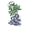
| ||||||||
|---|---|---|---|---|---|---|---|---|---|
| 1 | 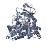
| ||||||||
| 2 | 
| ||||||||
| Unit cell |
|
-Components
-Protein , 1 types, 2 molecules AB
| #1: Protein | Mass: 46587.895 Da / Num. of mol.: 2 Source method: isolated from a genetically manipulated source Details: HEME ATTACHED VIA CYS357, XENON BOUND IN CAVITIES / Source: (gene. exp.) PSEUDOMONAS PUTIDA (bacteria) / Production host: |
|---|
-Non-polymers , 5 types, 467 molecules 


| #2: Chemical |  Mass: 616.487 Da / Num. of mol.: 2 / Source method: obtained synthetically / Formula: C34H32FeN4O4 Mass: 616.487 Da / Num. of mol.: 2 / Source method: obtained synthetically / Formula: C34H32FeN4O4#3: Chemical |  Mass: 152.233 Da / Num. of mol.: 2 / Source method: obtained synthetically / Formula: C10H16O Mass: 152.233 Da / Num. of mol.: 2 / Source method: obtained synthetically / Formula: C10H16O#4: Chemical |  Mass: 39.098 Da / Num. of mol.: 3 / Source method: obtained synthetically / Formula: K Mass: 39.098 Da / Num. of mol.: 3 / Source method: obtained synthetically / Formula: K#5: Chemical | ChemComp-XE /  Mass: 131.293 Da / Num. of mol.: 8 / Source method: obtained synthetically / Formula: Xe Mass: 131.293 Da / Num. of mol.: 8 / Source method: obtained synthetically / Formula: Xe#6: Water | ChemComp-HOH / | Mass: 18.015 Da / Num. of mol.: 452 / Source method: isolated from a natural source / Formula: H2O |
|---|
-Experimental details
-Experiment
| Experiment | Method: X-RAY DIFFRACTION / Number of used crystals: 1 |
|---|
- Sample preparation
| Crystal | Density Matthews: 2.14 Å3/Da / Density % sol: 42.55 % Description: A CRYOCOOLED, LOOP MOUNTED P450 CRYSTAL CONTAINING CAMPHOR AND HEME-BOUND CYANIDE (NO CYANIDE IN THE CRYOPROTECTANT CONTAINING 20% GLYCEROL) WAS WARMED UP QUICKLY AND MOUNTED IN A HOME- ...Description: A CRYOCOOLED, LOOP MOUNTED P450 CRYSTAL CONTAINING CAMPHOR AND HEME-BOUND CYANIDE (NO CYANIDE IN THE CRYOPROTECTANT CONTAINING 20% GLYCEROL) WAS WARMED UP QUICKLY AND MOUNTED IN A HOME-BUILT PRESSURE CELL. THE CRYSTAL WAS EXPOSED FOR 3 MIN. TO 7 BAR XENON AT 2 DEG. C SUBSEQUENTLY, THE CRYSTAL WAS FLASH COOLED IN LIQUID NITROGEN. |
|---|---|
| Crystal grow | Temperature: 275 K / Method: vapor diffusion, sitting drop / pH: 7.4 Details: CRYSTALS WERE GROWN USING THE SITTING DROP GEOMETRY AT 2 DEG. C. 5 UL OF 30 MG/ML P450 IN 50 MM TRIS HCL, 250 MM KCL, 0.5 MM CAMPHOR WERE MIXED WITH AN EQUAL VOLUME OF THE RESERVOIR SOLUTION ...Details: CRYSTALS WERE GROWN USING THE SITTING DROP GEOMETRY AT 2 DEG. C. 5 UL OF 30 MG/ML P450 IN 50 MM TRIS HCL, 250 MM KCL, 0.5 MM CAMPHOR WERE MIXED WITH AN EQUAL VOLUME OF THE RESERVOIR SOLUTION (27-30% PEG 4000, 100 MM DTE, SAME BUFFER AS PROTEIN)., pH 7.40 |
-Data collection
| Diffraction | Mean temperature: 100 K |
|---|---|
| Diffraction source | Source: SYNCHROTRON / Site: ESRF  / Type: ESRF / Wavelength: 0.934 / Type: ESRF / Wavelength: 0.934 |
| Detector | Type: ADSC CCD / Detector: CCD / Details: MIRRORS |
| Radiation | Monochromator: MONO / Protocol: SINGLE WAVELENGTH / Monochromatic (M) / Laue (L): M / Scattering type: x-ray |
| Radiation wavelength | Wavelength: 0.934 Å / Relative weight: 1 |
| Reflection | Resolution: 2→19 Å / Num. obs: 53282 / % possible obs: 99.7 % / Redundancy: 3.82 % / Biso Wilson estimate: 32.91 Å2 / Rmerge(I) obs: 0.121 / Net I/σ(I): 9.3 |
| Reflection shell | Resolution: 2→2.1 Å / Redundancy: 3.3 % / Rmerge(I) obs: 0.483 / Mean I/σ(I) obs: 4.2 / % possible all: 99.9 |
- Processing
| Software |
| ||||||||||||||||||||||||||||||||||||||||||||||||||||||||||||||||||||||||||||||||||||||||||||||||||||||||||||||||||||||||||||||||||||||||||||||||||||||||||||||||||||||||||||||||||||||
|---|---|---|---|---|---|---|---|---|---|---|---|---|---|---|---|---|---|---|---|---|---|---|---|---|---|---|---|---|---|---|---|---|---|---|---|---|---|---|---|---|---|---|---|---|---|---|---|---|---|---|---|---|---|---|---|---|---|---|---|---|---|---|---|---|---|---|---|---|---|---|---|---|---|---|---|---|---|---|---|---|---|---|---|---|---|---|---|---|---|---|---|---|---|---|---|---|---|---|---|---|---|---|---|---|---|---|---|---|---|---|---|---|---|---|---|---|---|---|---|---|---|---|---|---|---|---|---|---|---|---|---|---|---|---|---|---|---|---|---|---|---|---|---|---|---|---|---|---|---|---|---|---|---|---|---|---|---|---|---|---|---|---|---|---|---|---|---|---|---|---|---|---|---|---|---|---|---|---|---|---|---|---|---|
| Refinement | Method to determine structure: MOLECULAR REPLACEMENT Starting model: PDB ENTRY 1DZ8 Resolution: 2→19.84 Å / Cor.coef. Fo:Fc: 0.951 / Cor.coef. Fo:Fc free: 0.925 / SU B: 4.249 / SU ML: 0.119 / Cross valid method: THROUGHOUT / ESU R: 0.214 / ESU R Free: 0.179 / Stereochemistry target values: MAXIMUM LIKELIHOOD Details: HYDROGENS HAVE BEEN ADDED IN THE RIDING POSITIONS. N-TERMINUS IS DISORDERED
| ||||||||||||||||||||||||||||||||||||||||||||||||||||||||||||||||||||||||||||||||||||||||||||||||||||||||||||||||||||||||||||||||||||||||||||||||||||||||||||||||||||||||||||||||||||||
| Solvent computation | Ion probe radii: 0.8 Å / Shrinkage radii: 0.8 Å / VDW probe radii: 1.4 Å / Solvent model: BABINET MODEL WITH MASK | ||||||||||||||||||||||||||||||||||||||||||||||||||||||||||||||||||||||||||||||||||||||||||||||||||||||||||||||||||||||||||||||||||||||||||||||||||||||||||||||||||||||||||||||||||||||
| Displacement parameters | Biso mean: 31.9 Å2
| ||||||||||||||||||||||||||||||||||||||||||||||||||||||||||||||||||||||||||||||||||||||||||||||||||||||||||||||||||||||||||||||||||||||||||||||||||||||||||||||||||||||||||||||||||||||
| Refinement step | Cycle: LAST / Resolution: 2→19.84 Å
| ||||||||||||||||||||||||||||||||||||||||||||||||||||||||||||||||||||||||||||||||||||||||||||||||||||||||||||||||||||||||||||||||||||||||||||||||||||||||||||||||||||||||||||||||||||||
| Refine LS restraints |
|