 Movie
Movie Controller

[English] 日本語
 Yorodumi
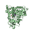
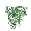
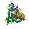
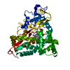
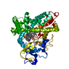
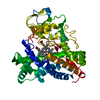
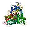
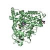
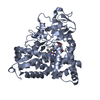
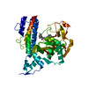
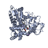
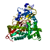
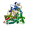
- PDB-1akd: CYTOCHROME P450CAM FROM PSEUDOMONAS PUTIDA, COMPLEXED WITH 1S-CAMPHOR -
+ Open data

- Basic information
| Entry | Database: PDB / ID: 1akd | ||||||
|---|---|---|---|---|---|---|---|
| Title | CYTOCHROME P450CAM FROM PSEUDOMONAS PUTIDA, COMPLEXED WITH 1S-CAMPHOR | ||||||
 Components Components | CYTOCHROME P450CAM | ||||||
 Keywords Keywords | OXIDOREDUCTASE / OXYGENASE / CYTOCHROME P450 / MONOOXYGENASE / ELECTRON TRANSPORT | ||||||
| Function / homology |  Function and homology information Function and homology informationcamphor 5-monooxygenase / camphor 5-monooxygenase activity / (+)-camphor catabolic process / iron ion binding / heme binding / cytoplasm Similarity search - Function | ||||||
| Biological species |  Pseudomonas putida (bacteria) Pseudomonas putida (bacteria) | ||||||
| Method |  X-RAY DIFFRACTION / SYNCHROTRON / MOLECULAR REPLACEMENT / Resolution: 1.8 Å X-RAY DIFFRACTION / SYNCHROTRON / MOLECULAR REPLACEMENT / Resolution: 1.8 Å | ||||||
 Authors Authors | Schlichting, I. / Jung, C. / Schulze, H. | ||||||
 Citation Citation | Journal: FEBS Lett. / Year: 1997 Title: Crystal structure of cytochrome P-450cam complexed with the (1S)-camphor enantiomer. Authors: Schlichting, I. / Jung, C. / Schulze, H. #1: Journal: J.Mol.Biol. / Year: 1987Title: High-Resolution Crystal Structure of Cytochrome P450Cam Authors: Poulos, T.L. / Finzel, B.C. / Howard, A.J. | ||||||
| History |
|
- Structure visualization
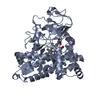
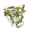
| Structure viewer | Molecule: MolmilJmol/JSmol |
|---|
- Downloads & links
-Download
| PDBx/mmCIF format | 1akd.cif.gz | 100.8 KB | Display | PDBx/mmCIF format |
|---|---|---|---|---|
| PDB format | pdb1akd.ent.gz | 75.7 KB | Display | PDB format |
| PDBx/mmJSON format | 1akd.json.gz | Tree view | PDBx/mmJSON format | |
| Others |  Other downloads Other downloads |
-Validation report
| Arichive directory | https://data.pdbj.org/pub/pdb/validation_reports/ak/1akdftp://data.pdbj.org/pub/pdb/validation_reports/ak/1akd | HTTPS FTP |
|---|
-Related structure data
| Similar structure data |
|---|


-Links
 PDBj
PDBj
- Assembly
| Deposited unit | 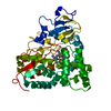
| ||||||||
|---|---|---|---|---|---|---|---|---|---|
| 1 |
| ||||||||
| Unit cell |
|
-Components
| #1: Protein | Mass: 46587.895 Da / Num. of mol.: 1 Source method: isolated from a genetically manipulated source Details: HEME LINKED BY CYS 357, SUBSTRATE (1S)-CAMPHOR BOUND IN TWO CONFORMATIONS, A AND B Source: (gene. exp.) Pseudomonas putida (bacteria) / Production host: |
|---|---|
| #2: Chemical | ChemComp-K /   Mass: 39.098 Da / Num. of mol.: 1 / Source method: obtained synthetically / Formula: K Mass: 39.098 Da / Num. of mol.: 1 / Source method: obtained synthetically / Formula: K |
| #3: Chemical | ChemComp-HEM /   Mass: 616.487 Da / Num. of mol.: 1 / Source method: obtained synthetically / Formula: C34H32FeN4O4 Mass: 616.487 Da / Num. of mol.: 1 / Source method: obtained synthetically / Formula: C34H32FeN4O4 |
| #4: Chemical | ChemComp-CAM / 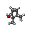  Mass: 152.233 Da / Num. of mol.: 1 / Source method: obtained synthetically / Formula: C10H16O Mass: 152.233 Da / Num. of mol.: 1 / Source method: obtained synthetically / Formula: C10H16O |
| #5: Water | ChemComp-HOH /  Mass: 18.015 Da / Num. of mol.: 271 / Source method: isolated from a natural source / Formula: H2O Mass: 18.015 Da / Num. of mol.: 271 / Source method: isolated from a natural source / Formula: H2O |
-Experimental details
-Experiment
| Experiment | Method: X-RAY DIFFRACTION / Number of used crystals: 1 |
|---|
- Sample preparation
| Crystal | Density Matthews: 2.44 Å3/Da / Density % sol: 49.55 % | ||||||||||||||||||||||||||||||||||||
|---|---|---|---|---|---|---|---|---|---|---|---|---|---|---|---|---|---|---|---|---|---|---|---|---|---|---|---|---|---|---|---|---|---|---|---|---|---|
| Crystal grow | Method: dialysis / pH: 7.4 Details: PROTEIN WAS CRYSTALLIZED BY DIALYSIS FROM 10% PEG 8000, 50 MM TRISHCL PH 7.4, 250 MM KCL, 100 MM DTE, IN THE PRESENCE OF EXCESS (1S)-CAMPHOR., dialysis | ||||||||||||||||||||||||||||||||||||
| Crystal grow | *PLUS Temperature: 4 ℃ / Method: unknown | ||||||||||||||||||||||||||||||||||||
| Components of the solutions | *PLUS
|
-Data collection
| Diffraction | Mean temperature: 277 K |
|---|---|
| Diffraction source | Source: SYNCHROTRON / Site: NSLS  / Beamline: X12C / Wavelength: 0.95 / Beamline: X12C / Wavelength: 0.95 |
| Detector | Type: MARRESEARCH / Detector: IMAGE PLATE AREA DETECTOR / Date: Jan 1, 1996 / Details: MIRRORS |
| Radiation | Monochromator: MONOCHROMATOR / Monochromatic (M) / Laue (L): M / Scattering type: x-ray |
| Radiation wavelength | Wavelength: 0.95 Å / Relative weight: 1 |
| Reflection | Resolution: 1.8→26 Å / Num. obs: 41095 / % possible obs: 92 % / Redundancy: 3.5 % / Rmerge(I) obs: 0.046 / Rsym value: 0.046 |
| Reflection shell | Resolution: 1.8→1.86 Å / Rmerge(I) obs: 0.284 / Rsym value: 0.284 / % possible all: 69.4 |
| Reflection | *PLUS Num. measured all: 143928 |
| Reflection shell | *PLUS % possible obs: 69.4 % |
- Processing
| Software |
| ||||||||||||||||||||||||||||||||||||||||||||||||||||||||||||
|---|---|---|---|---|---|---|---|---|---|---|---|---|---|---|---|---|---|---|---|---|---|---|---|---|---|---|---|---|---|---|---|---|---|---|---|---|---|---|---|---|---|---|---|---|---|---|---|---|---|---|---|---|---|---|---|---|---|---|---|---|---|
| Refinement | Method to determine structure: MOLECULAR REPLACEMENT Starting model: UNPUBLISHED COORDINATES FROM TETRAGONAL FORM OF P450CAM Resolution: 1.8→26 Å / Data cutoff high absF: 10000000 / Data cutoff low absF: 0
| ||||||||||||||||||||||||||||||||||||||||||||||||||||||||||||
| Displacement parameters | Biso mean: 29.7 Å2 | ||||||||||||||||||||||||||||||||||||||||||||||||||||||||||||
| Refinement step | Cycle: LAST / Resolution: 1.8→26 Å
| ||||||||||||||||||||||||||||||||||||||||||||||||||||||||||||
| Refine LS restraints |
| ||||||||||||||||||||||||||||||||||||||||||||||||||||||||||||
| LS refinement shell | Resolution: 1.8→1.88 Å / Total num. of bins used: 8
| ||||||||||||||||||||||||||||||||||||||||||||||||||||||||||||
| Xplor file |
| ||||||||||||||||||||||||||||||||||||||||||||||||||||||||||||
| Software | *PLUS Name: X-PLOR / Version: 3.851 / Classification: refinement | ||||||||||||||||||||||||||||||||||||||||||||||||||||||||||||
| Refine LS restraints | *PLUS
|
