 Movie
Movie Controller


+ Open data

- Basic information
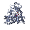
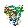
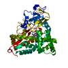
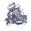
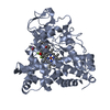
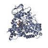
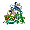
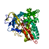
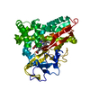
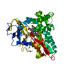
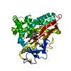
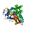
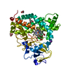
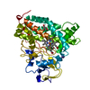
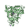
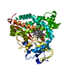
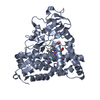
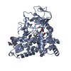
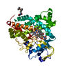
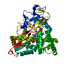
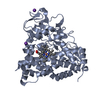
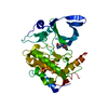
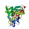
| Entry | Database: PDB / ID: 1o76 | ||||||
|---|---|---|---|---|---|---|---|
| Title | CYANIDE COMPLEX OF P450CAM FROM PSEUDOMONAS PUTIDA | ||||||
 Components Components | CYTOCHROME P450-CAM | ||||||
 Keywords Keywords | OXIDOREDUCTASE / MONO-OXYGENASE / HEME / ELECTRON TRANSPORT | ||||||
| Function / homology |  Function and homology information Function and homology informationcamphor 5-monooxygenase / camphor 5-monooxygenase activity / (+)-camphor catabolic process / iron ion binding / heme binding / cytoplasm Similarity search - Function | ||||||
| Biological species |  PSEUDOMONAS PUTIDA (bacteria) PSEUDOMONAS PUTIDA (bacteria) | ||||||
| Method |  X-RAY DIFFRACTION / SYNCHROTRON / FOURIER SYNTHESIS / Resolution: 1.8 Å X-RAY DIFFRACTION / SYNCHROTRON / FOURIER SYNTHESIS / Resolution: 1.8 Å | ||||||
 Authors Authors | Fedorov, R. / Ghosh, D. / Schlichting, I. | ||||||
 Citation Citation | Journal: Arch.Biochem.Biophys. / Year: 2003 Title: Crystal Structures of Cyanide Complexes of P450Cam and the Oxygenase Domain of Inducible Nitric Oxide Synthase-Structural Models of the Short-Lived Oxygen Complexes Authors: Fedorov, R. / Ghosh, D. / Schlichting, I. #1: Journal: Science / Year: 2000Title: The Catalytic Pathway of Cytochrome P450Cam at Atomic Resolution Authors: Schlichting, I. / Berendzen, J. / Chu, K. / Stock, A.M. / Maves, S.A. / Benson, D.E. / Sweet, R.M. / Ringe, D. / Petsko, G.A. / Sligar, S.G. #2: Journal: J.Mol.Biol. / Year: 1987Title: High-Resolution Crystal Structure of Cytochrome P450Cam Authors: Poulos, T.L. / Finzel, B.C. / Howard, A.J. | ||||||
| History |
|
- Structure visualization
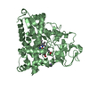
| Structure viewer | Molecule: MolmilJmol/JSmol |
|---|
- Downloads & links
-Download
| PDBx/mmCIF format | 1o76.cif.gz | 195.6 KB | Display | PDBx/mmCIF format |
|---|---|---|---|---|
| PDB format | pdb1o76.ent.gz | 155.5 KB | Display | PDB format |
| PDBx/mmJSON format | 1o76.json.gz | Tree view | PDBx/mmJSON format | |
| Others |  Other downloads Other downloads |
-Validation report
| Arichive directory | https://data.pdbj.org/pub/pdb/validation_reports/o7/1o76ftp://data.pdbj.org/pub/pdb/validation_reports/o7/1o76 | HTTPS FTP |
|---|
-Related structure data


-Links
 PDBj
PDBj


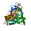
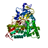
- Assembly
| Deposited unit | 
| ||||||||
|---|---|---|---|---|---|---|---|---|---|
| 1 | 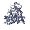
| ||||||||
| 2 | 
| ||||||||
| Unit cell |
| ||||||||
| Noncrystallographic symmetry (NCS) | NCS oper: (Code: given Matrix: (-0.9748, 0.0137, 0.2227), Vector: |
-Components
-Protein , 1 types, 2 molecules AB
| #1: Protein | Mass: 46587.895 Da / Num. of mol.: 2 Source method: isolated from a genetically manipulated source Details: CYANIDE BOUND TO HEME IRON. HEME ATTACHED VIA CYS357 Source: (gene. exp.) PSEUDOMONAS PUTIDA (bacteria) / Production host: |
|---|
-Non-polymers , 6 types, 961 molecules 

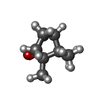


| #2: Chemical |  Mass: 616.487 Da / Num. of mol.: 2 / Source method: obtained synthetically / Formula: C34H32FeN4O4 Mass: 616.487 Da / Num. of mol.: 2 / Source method: obtained synthetically / Formula: C34H32FeN4O4#3: Chemical |  Mass: 26.017 Da / Num. of mol.: 2 / Source method: obtained synthetically / Formula: CN Mass: 26.017 Da / Num. of mol.: 2 / Source method: obtained synthetically / Formula: CN#4: Chemical |  Mass: 152.233 Da / Num. of mol.: 2 / Source method: obtained synthetically / Formula: C10H16O Mass: 152.233 Da / Num. of mol.: 2 / Source method: obtained synthetically / Formula: C10H16O#5: Chemical | ChemComp-TRS / |  Mass: 122.143 Da / Num. of mol.: 1 / Source method: obtained synthetically / Formula: C4H12NO3 / Comment: pH buffer*YM Mass: 122.143 Da / Num. of mol.: 1 / Source method: obtained synthetically / Formula: C4H12NO3 / Comment: pH buffer*YM#6: Chemical |  Mass: 39.098 Da / Num. of mol.: 3 / Source method: obtained synthetically / Formula: K Mass: 39.098 Da / Num. of mol.: 3 / Source method: obtained synthetically / Formula: K#7: Water | ChemComp-HOH / | Mass: 18.015 Da / Num. of mol.: 951 / Source method: isolated from a natural source / Formula: H2O |
|---|
-Experimental details
-Experiment
| Experiment | Method: X-RAY DIFFRACTION / Number of used crystals: 1 |
|---|
- Sample preparation
| Crystal | Density Matthews: 1.87 Å3/Da / Density % sol: 33.71 % | |||||||||||||||||||||||||||||||||||||||||||||||||||||||||||||||||||||||||||||||||||||||||||
|---|---|---|---|---|---|---|---|---|---|---|---|---|---|---|---|---|---|---|---|---|---|---|---|---|---|---|---|---|---|---|---|---|---|---|---|---|---|---|---|---|---|---|---|---|---|---|---|---|---|---|---|---|---|---|---|---|---|---|---|---|---|---|---|---|---|---|---|---|---|---|---|---|---|---|---|---|---|---|---|---|---|---|---|---|---|---|---|---|---|---|---|---|
| Crystal grow | Temperature: 275 K / Method: vapor diffusion, sitting drop / pH: 7.4 Details: CRYSTALS WERE GROWN USING THE SITTING DROP GEOMETRY AT 2 DEG. C. 5 UL OF 30 MG/ML P450 IN 50 MM TRIS HCL, 250 MM KCL, 0.5 MM CAMPHOR WERE MIXED WITH AN EQUAL VOLUME OF THE RESERVOIR SOLUTION ...Details: CRYSTALS WERE GROWN USING THE SITTING DROP GEOMETRY AT 2 DEG. C. 5 UL OF 30 MG/ML P450 IN 50 MM TRIS HCL, 250 MM KCL, 0.5 MM CAMPHOR WERE MIXED WITH AN EQUAL VOLUME OF THE RESERVOIR SOLUTION (27-30% PEG 4000, 100 MM DTE, SAME BUFFER AS PROTEIN)., pH 7.40 | |||||||||||||||||||||||||||||||||||||||||||||||||||||||||||||||||||||||||||||||||||||||||||
| Crystal grow | *PLUS Temperature: 4 ℃ / pH: 7.6 / Method: vapor diffusion, hanging drop | |||||||||||||||||||||||||||||||||||||||||||||||||||||||||||||||||||||||||||||||||||||||||||
| Components of the solutions | *PLUS
|
-Data collection
| Diffraction | Mean temperature: 100 K |
|---|---|
| Diffraction source | Source: SYNCHROTRON / Site: NSLS  / Beamline: X12C / Wavelength: 1 / Beamline: X12C / Wavelength: 1 |
| Detector | Type: BRANDEIS / Detector: CCD |
| Radiation | Protocol: SINGLE WAVELENGTH / Monochromatic (M) / Laue (L): M / Scattering type: x-ray |
| Radiation wavelength | Wavelength: 1 Å / Relative weight: 1 |
| Reflection | Resolution: 1.8→20 Å / Num. obs: 67892 / % possible obs: 94.5 % / Redundancy: 3.49 % / Biso Wilson estimate: 27 Å2 / Rsym value: 0.082 / Net I/σ(I): 8.2 |
| Reflection shell | Resolution: 1.8→2 Å / Redundancy: 2.8 % / Mean I/σ(I) obs: 4 / Rsym value: 0.242 / % possible all: 88.9 |
| Reflection | *PLUS Highest resolution: 1.8 Å / Num. measured all: 237007 / Rmerge(I) obs: 0.082 |
| Reflection shell | *PLUS % possible obs: 88.9 % / Rmerge(I) obs: 0.242 |
- Processing
| Software |
| ||||||||||||||||||||||||||||||||||||||||||||||||||||||||||||
|---|---|---|---|---|---|---|---|---|---|---|---|---|---|---|---|---|---|---|---|---|---|---|---|---|---|---|---|---|---|---|---|---|---|---|---|---|---|---|---|---|---|---|---|---|---|---|---|---|---|---|---|---|---|---|---|---|---|---|---|---|---|
| Refinement | Method to determine structure: FOURIER SYNTHESIS / Resolution: 1.8→19 Å / Data cutoff high absF: 10000000 / Cross valid method: THROUGHOUT / σ(F): 0 / Details: N-TERMINUS IS DISORDERED
| ||||||||||||||||||||||||||||||||||||||||||||||||||||||||||||
| Solvent computation | Bsol: 36.16 Å2 | ||||||||||||||||||||||||||||||||||||||||||||||||||||||||||||
| Refinement step | Cycle: LAST / Resolution: 1.8→19 Å
| ||||||||||||||||||||||||||||||||||||||||||||||||||||||||||||
| Refine LS restraints |
| ||||||||||||||||||||||||||||||||||||||||||||||||||||||||||||
| Refinement | *PLUS Lowest resolution: 20 Å / % reflection Rfree: 5 % / Rfactor Rfree: 0.236 / Rfactor Rwork: 0.196 | ||||||||||||||||||||||||||||||||||||||||||||||||||||||||||||
| Solvent computation | *PLUS | ||||||||||||||||||||||||||||||||||||||||||||||||||||||||||||
| Displacement parameters | *PLUS | ||||||||||||||||||||||||||||||||||||||||||||||||||||||||||||
| Refine LS restraints | *PLUS
|
