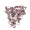
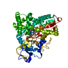
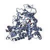
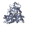
 Movie
Movie Controller

[English] 日本語
 Yorodumi
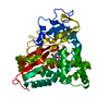
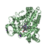
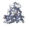
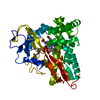
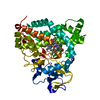
- PDB-2zaw: Crystal Structure of Ferric Cytochrome P450cam Reconstituted with... -
+ Open data

- Basic information
| Entry | Database: PDB / ID: 2zaw | ||||||
|---|---|---|---|---|---|---|---|
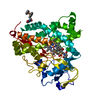
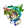
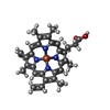
| Title | Crystal Structure of Ferric Cytochrome P450cam Reconstituted with 6-Methyl-6-depropionated Hemin | ||||||
 Components Components | Cytochrome P450-cam | ||||||
 Keywords Keywords | OXIDOREDUCTASE / P450CAM / MONOOXYGENASE / CAMPHOR-HYDROXYLASE / Heme / Iron / Membrane / Metal-binding | ||||||
| Function / homology |  Function and homology information Function and homology informationcamphor 5-monooxygenase / camphor 5-monooxygenase activity / (+)-camphor catabolic process / iron ion binding / heme binding / cytoplasm Similarity search - Function | ||||||
| Biological species |  Pseudomonas putida (bacteria) Pseudomonas putida (bacteria) | ||||||
| Method |  X-RAY DIFFRACTION / SYNCHROTRON / MOLECULAR REPLACEMENT / Resolution: 1.55 Å X-RAY DIFFRACTION / SYNCHROTRON / MOLECULAR REPLACEMENT / Resolution: 1.55 Å | ||||||
 Authors Authors | Harada, K. / Sakurai, K. / Shimada, H. / Tsukihara, T. / Hayashi, T. | ||||||
 Citation Citation | Journal: J.Am.Chem.Soc. / Year: 2008 Title: Evaluation of the functional role of the heme-6-propionate side chain in cytochrome P450cam Authors: Harada, K. / Sakurai, K. / Ikemura, K. / Ogura, T. / Hirota, S. / Shimada, H. / Hayashi, T. | ||||||
| History |
|
- Structure visualization
| Structure viewer | Molecule: MolmilJmol/JSmol |
|---|
- Downloads & links
-Download
| PDBx/mmCIF format | 2zaw.cif.gz | 122.8 KB | Display | PDBx/mmCIF format |
|---|---|---|---|---|
| PDB format | pdb2zaw.ent.gz | 91.4 KB | Display | PDB format |
| PDBx/mmJSON format | 2zaw.json.gz | Tree view | PDBx/mmJSON format | |
| Others |  Other downloads Other downloads |
-Validation report
| Arichive directory | https://data.pdbj.org/pub/pdb/validation_reports/za/2zawftp://data.pdbj.org/pub/pdb/validation_reports/za/2zaw | HTTPS FTP |
|---|
-Related structure data
| Related structure data |  2zaxC  2cppS C: citing same article ( S: Starting model for refinement |
|---|---|
| Similar structure data |


-Links
 PDBj
PDBj
- Assembly
| Deposited unit | 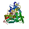
| ||||||||
|---|---|---|---|---|---|---|---|---|---|
| 1 |
| ||||||||
| Unit cell |
|
-Components
-Protein , 1 types, 1 molecules A
| #1: Protein | Mass: 46720.074 Da / Num. of mol.: 1 Source method: isolated from a genetically manipulated source Source: (gene. exp.) Pseudomonas putida (bacteria) / Gene: camC, cyp101 / Plasmid: M13tv19 / Production host: |
|---|
-Non-polymers , 5 types, 800 molecules 


| #2: Chemical | ChemComp-K /  Mass: 39.098 Da / Num. of mol.: 1 / Source method: obtained synthetically / Formula: K Mass: 39.098 Da / Num. of mol.: 1 / Source method: obtained synthetically / Formula: K |
|---|---|
| #3: Chemical | ChemComp-CL /  Mass: 35.453 Da / Num. of mol.: 1 / Source method: obtained synthetically / Formula: Cl Mass: 35.453 Da / Num. of mol.: 1 / Source method: obtained synthetically / Formula: Cl |
| #4: Chemical | ChemComp-6HE /  Mass: 558.451 Da / Num. of mol.: 1 / Source method: obtained synthetically / Formula: C32H30FeN4O2 Mass: 558.451 Da / Num. of mol.: 1 / Source method: obtained synthetically / Formula: C32H30FeN4O2 |
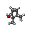
| #5: Chemical | ChemComp-CAM /  Mass: 152.233 Da / Num. of mol.: 1 / Source method: obtained synthetically / Formula: C10H16O Mass: 152.233 Da / Num. of mol.: 1 / Source method: obtained synthetically / Formula: C10H16O |
| #6: Water | ChemComp-HOH / Mass: 18.015 Da / Num. of mol.: 796 / Source method: isolated from a natural source / Formula: H2O |
-Experimental details
-Experiment
| Experiment | Method: X-RAY DIFFRACTION / Number of used crystals: 1 |
|---|
- Sample preparation
| Crystal | Density Matthews: 2.71 Å3/Da / Density % sol: 54.69 % |
|---|---|
| Crystal grow | Temperature: 298 K / Method: vapor diffusion, sitting drop / pH: 7.4 Details: 14% PEG 8000, 50mM Tris-HCl, 250mM KCl, 1mM d-camphor, 10mM dithioerythritol(DTE), pH 7.4, VAPOR DIFFUSION, SITTING DROP, temperature 298K |
-Data collection
| Diffraction | Mean temperature: 100 K |
|---|---|
| Diffraction source | Source: SYNCHROTRON / Site: SPring-8  / Beamline: BL44XU / Wavelength: 0.7 Å / Beamline: BL44XU / Wavelength: 0.7 Å |
| Detector | Type: Bruker DIP-6040 / Detector: CCD / Date: Jun 11, 2007 |
| Radiation | Monochromator: Si / Protocol: SINGLE WAVELENGTH / Monochromatic (M) / Laue (L): M / Scattering type: x-ray |
| Radiation wavelength | Wavelength: 0.7 Å / Relative weight: 1 |
| Reflection | Resolution: 1.55→45.04 Å / Num. all: 75813 / Num. obs: 75813 / % possible obs: 99.8 % / Redundancy: 7.3 % / Biso Wilson estimate: 13.92 Å2 / Rmerge(I) obs: 0.07 / Net I/σ(I): 9.1 |
| Reflection shell | Resolution: 1.55→1.59 Å / Redundancy: 7.4 % / Rmerge(I) obs: 0.387 / Mean I/σ(I) obs: 5.9 / Num. unique all: 4929 / % possible all: 100 |
- Processing
| Software |
| |||||||||||||||||||||||||||||||||||||||||||||||||||||||||||||||||||||||||||||||||||||||||||||||
|---|---|---|---|---|---|---|---|---|---|---|---|---|---|---|---|---|---|---|---|---|---|---|---|---|---|---|---|---|---|---|---|---|---|---|---|---|---|---|---|---|---|---|---|---|---|---|---|---|---|---|---|---|---|---|---|---|---|---|---|---|---|---|---|---|---|---|---|---|---|---|---|---|---|---|---|---|---|---|---|---|---|---|---|---|---|---|---|---|---|---|---|---|---|---|---|---|
| Refinement | Method to determine structure: MOLECULAR REPLACEMENT Starting model: PDB ENTRY 2CPP Resolution: 1.55→45.04 Å / Cor.coef. Fo:Fc: 0.969 / Cor.coef. Fo:Fc free: 0.957 / SU B: 1.11 / SU ML: 0.041 / Isotropic thermal model: mixed / Cross valid method: THROUGHOUT / ESU R: 0.069 / ESU R Free: 0.07 / Stereochemistry target values: MAXIMUM LIKELIHOOD / Details: HYDROGENS HAVE BEEN ADDED IN THE RIDING POSITIONS
| |||||||||||||||||||||||||||||||||||||||||||||||||||||||||||||||||||||||||||||||||||||||||||||||
| Solvent computation | Ion probe radii: 0.8 Å / Shrinkage radii: 0.8 Å / VDW probe radii: 1.2 Å / Solvent model: MASK | |||||||||||||||||||||||||||||||||||||||||||||||||||||||||||||||||||||||||||||||||||||||||||||||
| Displacement parameters | Biso mean: 16.417 Å2
| |||||||||||||||||||||||||||||||||||||||||||||||||||||||||||||||||||||||||||||||||||||||||||||||
| Refinement step | Cycle: LAST / Resolution: 1.55→45.04 Å
| |||||||||||||||||||||||||||||||||||||||||||||||||||||||||||||||||||||||||||||||||||||||||||||||
| Refine LS restraints |
| |||||||||||||||||||||||||||||||||||||||||||||||||||||||||||||||||||||||||||||||||||||||||||||||
| LS refinement shell | Resolution: 1.551→1.591 Å / Total num. of bins used: 20
|
