 Movie
Movie Controller

[English] 日本語
 Yorodumi
- PDB-2qbo: Crystal structure of the P450cam G248V mutant in the cyanide boun... -
+ Open data

- Basic information
| Entry | Database: PDB / ID: 2qbo | ||||||
|---|---|---|---|---|---|---|---|
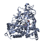
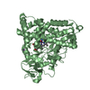
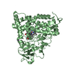
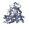
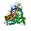
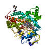
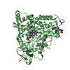
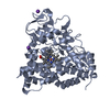
| Title | Crystal structure of the P450cam G248V mutant in the cyanide bound state | ||||||
 Components Components | Cytochrome P450-cam | ||||||
 Keywords Keywords | OXIDOREDUCTASE / CYP101 / mutant / conserved active site residue / cyanide complex / Gly248 / heme geometry | ||||||
| Function / homology |  Function and homology information Function and homology informationcamphor 5-monooxygenase / camphor 5-monooxygenase activity / (+)-camphor catabolic process / iron ion binding / heme binding / cytoplasm Similarity search - Function | ||||||
| Biological species |  Pseudomonas putida (bacteria) Pseudomonas putida (bacteria) | ||||||
| Method |  X-RAY DIFFRACTION / SYNCHROTRON / FOURIER SYNTHESIS / Resolution: 1.9 Å X-RAY DIFFRACTION / SYNCHROTRON / FOURIER SYNTHESIS / Resolution: 1.9 Å | ||||||
 Authors Authors | von Koenig, K. / Makris, T.M. / Sligar, S.D. / Schlichting, I. | ||||||
 Citation Citation | Journal: Biochemistry / Year: 2007 Title: Alteration of P450 Distal Pocket Solvent Leads to Impaired Proton Delivery and Changes in Heme Geometry. Authors: Makris, T.M. / Koenig, K.V. / Schlichting, I. / Sligar, S.G. | ||||||
| History |
|
- Structure visualization
| Structure viewer | Molecule: MolmilJmol/JSmol |
|---|
- Downloads & links
-Download
| PDBx/mmCIF format | 2qbo.cif.gz | 109.1 KB | Display | PDBx/mmCIF format |
|---|---|---|---|---|
| PDB format | pdb2qbo.ent.gz | 80.9 KB | Display | PDB format |
| PDBx/mmJSON format | 2qbo.json.gz | Tree view | PDBx/mmJSON format | |
| Others |  Other downloads Other downloads |
-Validation report
| Arichive directory | https://data.pdbj.org/pub/pdb/validation_reports/qb/2qboftp://data.pdbj.org/pub/pdb/validation_reports/qb/2qbo | HTTPS FTP |
|---|
-Related structure data
| Related structure data |  2qblC  2qbmC  2qbnC  1yrcS C: citing same article ( S: Starting model for refinement |
|---|---|
| Similar structure data |


-Links
 PDBj
PDBj


- Assembly
| Deposited unit | 
| ||||||||
|---|---|---|---|---|---|---|---|---|---|
| 1 |
| ||||||||
| Unit cell |
| ||||||||
| Components on special symmetry positions |
|
-Components
-Protein , 1 types, 1 molecules A
| #1: Protein | Mass: 47591.023 Da / Num. of mol.: 1 / Mutation: G248V Source method: isolated from a genetically manipulated source Source: (gene. exp.) Pseudomonas putida (bacteria) / Gene: camC, cyp101 / Plasmid: pET28 / Species (production host): Escherichia coli / Production host: |
|---|
-Non-polymers , 5 types, 524 molecules 


| #2: Chemical | ChemComp-CYN /  Mass: 26.017 Da / Num. of mol.: 1 / Source method: obtained synthetically / Formula: CN Mass: 26.017 Da / Num. of mol.: 1 / Source method: obtained synthetically / Formula: CN |
|---|---|
| #3: Chemical | ChemComp-K /  Mass: 39.098 Da / Num. of mol.: 1 / Source method: obtained synthetically / Formula: K Mass: 39.098 Da / Num. of mol.: 1 / Source method: obtained synthetically / Formula: K |
| #4: Chemical | ChemComp-HEM /  Mass: 616.487 Da / Num. of mol.: 1 / Source method: obtained synthetically / Formula: C34H32FeN4O4 Mass: 616.487 Da / Num. of mol.: 1 / Source method: obtained synthetically / Formula: C34H32FeN4O4 |
| #5: Chemical | ChemComp-CAM /  Mass: 152.233 Da / Num. of mol.: 1 / Source method: obtained synthetically / Formula: C10H16O Mass: 152.233 Da / Num. of mol.: 1 / Source method: obtained synthetically / Formula: C10H16O |
| #6: Water | ChemComp-HOH / Mass: 18.015 Da / Num. of mol.: 520 / Source method: isolated from a natural source / Formula: H2O |
-Experimental details
-Experiment
| Experiment | Method: X-RAY DIFFRACTION / Number of used crystals: 1 |
|---|
- Sample preparation
| Crystal | Density Matthews: 2.57 Å3/Da / Density % sol: 52.1 % |
|---|---|
| Crystal grow | Temperature: 277 K / Method: vapor diffusion, sitting drop / pH: 7.4 Details: 1 UL OF 15 MG/ML P450 IN 50 MM Potassium phosphate, 250 MM KCL WERE MIXED WITH AN EQUAL VOLUME OF THE RESERVOIR SOLUTION (100 mM Tris, 250 mM KCl, 27-30% PEG 8000, 100 MM DTE), pH 7.4, VAPOR ...Details: 1 UL OF 15 MG/ML P450 IN 50 MM Potassium phosphate, 250 MM KCL WERE MIXED WITH AN EQUAL VOLUME OF THE RESERVOIR SOLUTION (100 mM Tris, 250 mM KCl, 27-30% PEG 8000, 100 MM DTE), pH 7.4, VAPOR DIFFUSION, SITTING DROP, temperature 277K |
-Data collection
| Diffraction | Mean temperature: 100 K |
|---|---|
| Diffraction source | Source: SYNCHROTRON / Site: SLS  / Beamline: X10SA / Wavelength: 1.006 Å / Beamline: X10SA / Wavelength: 1.006 Å |
| Detector | Type: MARRESEARCH / Detector: CCD / Date: Nov 12, 2005 / Details: DYNAMICALLY BENDABLE MIRROR |
| Radiation | Monochromator: SI(111) / Protocol: SINGLE WAVELENGTH / Monochromatic (M) / Laue (L): M / Scattering type: x-ray |
| Radiation wavelength | Wavelength: 1.006 Å / Relative weight: 1 |
| Reflection | Resolution: 1.9→20 Å / Num. all: 39886 / Num. obs: 39886 / % possible obs: 99 % / Observed criterion σ(I): -3 / Redundancy: 3.9 % / Rsym value: 0.049 / Net I/σ(I): 15 |
| Reflection shell | Resolution: 1.9→2 Å / Redundancy: 3.1 % / Mean I/σ(I) obs: 3.3 / Num. unique all: 5399 / Rsym value: 0.334 / % possible all: 96 |
- Processing
| Software |
| ||||||||||||||||||||||||||||
|---|---|---|---|---|---|---|---|---|---|---|---|---|---|---|---|---|---|---|---|---|---|---|---|---|---|---|---|---|---|
| Refinement | Method to determine structure: FOURIER SYNTHESIS Starting model: PDB entry 1yrc Resolution: 1.9→20 Å / Cross valid method: THROUGHOUT / σ(F): 0 / Stereochemistry target values: maximum likelihood
| ||||||||||||||||||||||||||||
| Solvent computation | Bsol: 39.019 Å2 | ||||||||||||||||||||||||||||
| Displacement parameters | Biso mean: 33.067 Å2
| ||||||||||||||||||||||||||||
| Refinement step | Cycle: LAST / Resolution: 1.9→20 Å
| ||||||||||||||||||||||||||||
| Refine LS restraints |
| ||||||||||||||||||||||||||||
| Xplor file |
|
