 Movie
Movie Controller


+ Open data

- Basic information
| Entry | Database: PDB / ID: 1hjo | ||||||
|---|---|---|---|---|---|---|---|
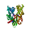
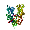
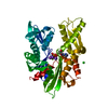
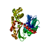
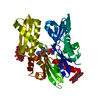
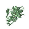
| Title | ATPase domain of human heat shock 70kDa protein 1 | ||||||
 Components Components | PROTEIN (HEAT-SHOCK 70KD PROTEIN) | ||||||
 Keywords Keywords | HYDROLASE / ATP-BINDING / CHAPERONE / HEAT SHOCK | ||||||
| Function / homology |  Function and homology information Function and homology information: / : / : / : / cellular heat acclimation / negative regulation of inclusion body assembly / positive regulation of nucleotide-binding oligomerization domain containing 2 signaling pathway / Viral RNP Complexes in the Host Cell Nucleus / C3HC4-type RING finger domain binding / positive regulation of microtubule nucleation ...: / : / : / : / cellular heat acclimation / negative regulation of inclusion body assembly / positive regulation of nucleotide-binding oligomerization domain containing 2 signaling pathway / Viral RNP Complexes in the Host Cell Nucleus / C3HC4-type RING finger domain binding / positive regulation of microtubule nucleation / ATP-dependent protein disaggregase activity / misfolded protein binding / negative regulation of mitochondrial outer membrane permeabilization involved in apoptotic signaling pathway / regulation of mitotic spindle assembly / positive regulation of tumor necrosis factor-mediated signaling pathway / aggresome / lysosomal transport / cellular response to steroid hormone stimulus / mRNA catabolic process / regulation of protein ubiquitination / cellular response to unfolded protein / HSF1-dependent transactivation / Regulation of HSF1-mediated heat shock response / response to unfolded protein / negative regulation of extrinsic apoptotic signaling pathway in absence of ligand / Mitochondrial unfolded protein response (UPRmt) / Attenuation phase / negative regulation of endoplasmic reticulum stress-induced intrinsic apoptotic signaling pathway / chaperone-mediated protein complex assembly / transcription regulator inhibitor activity / ATP metabolic process / vesicle-mediated transport / endoplasmic reticulum unfolded protein response / heat shock protein binding / : / protein folding chaperone / centriole / inclusion body / negative regulation of protein ubiquitination / positive regulation of erythrocyte differentiation / HSP90 chaperone cycle for steroid hormone receptors (SHR) in the presence of ligand / positive regulation of RNA splicing / positive regulation of interleukin-8 production / ATP-dependent protein folding chaperone / negative regulation of transforming growth factor beta receptor signaling pathway / AUF1 (hnRNP D0) binds and destabilizes mRNA / negative regulation of cell growth / PKR-mediated signaling / G protein-coupled receptor binding / histone deacetylase binding / disordered domain specific binding / : / transcription corepressor activity / positive regulation of proteasomal ubiquitin-dependent protein catabolic process / cellular response to heat / protein refolding / virus receptor activity / cellular response to oxidative stress / blood microparticle / vesicle / ficolin-1-rich granule lumen / nuclear speck / protein stabilization / cadherin binding / ribonucleoprotein complex / receptor ligand activity / negative regulation of cell population proliferation / signaling receptor binding / focal adhesion / centrosome / positive regulation of gene expression / ubiquitin protein ligase binding / Neutrophil degranulation / negative regulation of apoptotic process / perinuclear region of cytoplasm / enzyme binding / negative regulation of transcription by RNA polymerase II / endoplasmic reticulum / ATP hydrolysis activity / protein-containing complex / mitochondrion / : / RNA binding / extracellular exosome / extracellular region / nucleoplasm / ATP binding / nucleus / plasma membrane / cytosol / cytoplasm Similarity search - Function | ||||||
| Biological species |  Homo sapiens (human) Homo sapiens (human) | ||||||
| Method |  X-RAY DIFFRACTION / SYNCHROTRON / MOLECULAR REPLACEMENT / Resolution: 2.3 Å X-RAY DIFFRACTION / SYNCHROTRON / MOLECULAR REPLACEMENT / Resolution: 2.3 Å | ||||||
 Authors Authors | Osipiuk, J. / Walsh, M.A. / Freeman, B.C. / Morimoto, R.I. / Joachimiak, A. | ||||||
 Citation Citation | Journal: Acta Crystallogr.,Sect.D / Year: 1999 Title: Structure of a new crystal form of human Hsp70 ATPase domain. Authors: Osipiuk, J. / Walsh, M.A. / Freeman, B.C. / Morimoto, R.I. / Joachimiak, A. #1: Journal: Structure / Year: 1997Title: Human Hsp70 Molecular Chaperone Binds Two Calcium Ions within the ATPase Domain Authors: Sriram, M. / Osipiuk, J. / Freeman, B.C. / Morimoto, R.I. / Joachimiak, A. #2: Journal: Nature / Year: 1990Title: Three-Dimensional Structure of the ATPase Fragment of a 70K Heat-Shock Cognate Protein Authors: Flaherty, K.M. / Deluca-Flaherty, C. / Mckay, D.B. | ||||||
| History |
|
- Structure visualization
| Structure viewer | Molecule: MolmilJmol/JSmol |
|---|
- Downloads & links
-Download
| PDBx/mmCIF format | 1hjo.cif.gz | 91.8 KB | Display | PDBx/mmCIF format |
|---|---|---|---|---|
| PDB format | pdb1hjo.ent.gz | 68.2 KB | Display | PDB format |
| PDBx/mmJSON format | 1hjo.json.gz | Tree view | PDBx/mmJSON format | |
| Others |  Other downloads Other downloads |
-Validation report
| Arichive directory | https://data.pdbj.org/pub/pdb/validation_reports/hj/1hjoftp://data.pdbj.org/pub/pdb/validation_reports/hj/1hjo | HTTPS FTP |
|---|
-Related structure data
| Similar structure data |
|---|


-Links
 PDBj
PDBj


- Assembly
| Deposited unit | 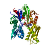
| ||||||||
|---|---|---|---|---|---|---|---|---|---|
| 1 |
| ||||||||
| Unit cell |
|
-Components
| #1: Protein | Mass: 41882.387 Da / Num. of mol.: 1 / Fragment: 42KD ATPASE N-TERMINAL FRAGMENT Source method: isolated from a genetically manipulated source Source: (gene. exp.) Homo sapiens (human) / Cellular location: CYTOSOL / Cell line (production host): BL21(DE3) / Production host:  References: UniProt: P08107, UniProt: P0DMV8*PLUS, EC: 3.6.1.3 |
|---|---|
| #2: Chemical | ChemComp-CA /   Mass: 40.078 Da / Num. of mol.: 1 / Source method: obtained synthetically / Formula: Ca Mass: 40.078 Da / Num. of mol.: 1 / Source method: obtained synthetically / Formula: Ca |
| #3: Chemical | ChemComp-CL /   Mass: 35.453 Da / Num. of mol.: 1 / Source method: obtained synthetically / Formula: Cl Mass: 35.453 Da / Num. of mol.: 1 / Source method: obtained synthetically / Formula: Cl |
| #4: Chemical | ChemComp-ADP /   Mass: 427.201 Da / Num. of mol.: 1 / Source method: obtained synthetically / Formula: C10H15N5O10P2 / Comment: ADP, energy-carrying molecule*YM Mass: 427.201 Da / Num. of mol.: 1 / Source method: obtained synthetically / Formula: C10H15N5O10P2 / Comment: ADP, energy-carrying molecule*YM |
| #5: Water | ChemComp-HOH /  Mass: 18.015 Da / Num. of mol.: 199 / Source method: isolated from a natural source / Formula: H2O Mass: 18.015 Da / Num. of mol.: 199 / Source method: isolated from a natural source / Formula: H2O |
-Experimental details
-Experiment
| Experiment | Method: X-RAY DIFFRACTION / Number of used crystals: 1 |
|---|
- Sample preparation
| Crystal | Density Matthews: 3.01 Å3/Da / Density % sol: 59.11 % Description: MODEL USED WAS UNSUBMITTED REFINED HUMAN HSP70 ATPASE | ||||||||||||||||||||||||||||||||||||
|---|---|---|---|---|---|---|---|---|---|---|---|---|---|---|---|---|---|---|---|---|---|---|---|---|---|---|---|---|---|---|---|---|---|---|---|---|---|
| Crystal grow | pH: 7 Details: 8% PEG-8000 25 MM IMIDAZOLE, PH 7.0 20MM CACL2 1MM GAMMA-S-ATP | ||||||||||||||||||||||||||||||||||||
| Crystal grow | *PLUS Method: vapor diffusion, hanging drop | ||||||||||||||||||||||||||||||||||||
| Components of the solutions | *PLUS
|
-Data collection
| Diffraction | Mean temperature: 120 K |
|---|---|
| Diffraction source | Source: SYNCHROTRON / Site: NSLS  / Beamline: X12B / Wavelength: 1 / Beamline: X12B / Wavelength: 1 |
| Detector | Type: MARRESEARCH / Detector: IMAGE PLATE / Date: Jan 15, 1996 |
| Radiation | Protocol: SINGLE WAVELENGTH / Monochromatic (M) / Laue (L): M / Scattering type: x-ray |
| Radiation wavelength | Wavelength: 1 Å / Relative weight: 1 |
| Reflection | Resolution: 2.2→10 Å / Num. obs: 20763 / % possible obs: 90.7 % / Observed criterion σ(I): 3 / Redundancy: 2.7 % / Biso Wilson estimate: 24.6 Å2 / Rmerge(I) obs: 0.05 / Net I/σ(I): 22.1 |
| Reflection shell | Resolution: 2.3→2.34 Å / Rmerge(I) obs: 0.205 / Mean I/σ(I) obs: 5.5 / % possible all: 80.7 |
| Reflection | *PLUS Num. measured all: 56187 |
| Reflection shell | *PLUS % possible obs: 80.7 % |
- Processing
| Software |
| ||||||||||||||||||||||||||||||||||||||||||||||||||||||||||||||||||||||||||||||||
|---|---|---|---|---|---|---|---|---|---|---|---|---|---|---|---|---|---|---|---|---|---|---|---|---|---|---|---|---|---|---|---|---|---|---|---|---|---|---|---|---|---|---|---|---|---|---|---|---|---|---|---|---|---|---|---|---|---|---|---|---|---|---|---|---|---|---|---|---|---|---|---|---|---|---|---|---|---|---|---|---|---|
| Refinement | Method to determine structure: MOLECULAR REPLACEMENT / Resolution: 2.3→9 Å / Rfactor Rfree error: 0.006 / Data cutoff high rms absF: 265761.08 / Isotropic thermal model: RESTRAINED / Cross valid method: THROUGHOUT / σ(F): 1.5
| ||||||||||||||||||||||||||||||||||||||||||||||||||||||||||||||||||||||||||||||||
| Solvent computation | Solvent model: FLAT MODEL / Bsol: 59.74 Å2 / ksol: 0.448 e/Å3 | ||||||||||||||||||||||||||||||||||||||||||||||||||||||||||||||||||||||||||||||||
| Displacement parameters | Biso mean: 30.4 Å2 | ||||||||||||||||||||||||||||||||||||||||||||||||||||||||||||||||||||||||||||||||
| Refine analyze |
| ||||||||||||||||||||||||||||||||||||||||||||||||||||||||||||||||||||||||||||||||
| Refinement step | Cycle: LAST / Resolution: 2.3→9 Å
| ||||||||||||||||||||||||||||||||||||||||||||||||||||||||||||||||||||||||||||||||
| Refine LS restraints |
| ||||||||||||||||||||||||||||||||||||||||||||||||||||||||||||||||||||||||||||||||
| LS refinement shell | Resolution: 2.3→2.44 Å / Rfactor Rfree error: 0.016 / Total num. of bins used: 6
| ||||||||||||||||||||||||||||||||||||||||||||||||||||||||||||||||||||||||||||||||
| Xplor file |
| ||||||||||||||||||||||||||||||||||||||||||||||||||||||||||||||||||||||||||||||||
| Software | *PLUS Name: CNS / Version: 0.4 / Classification: refinement | ||||||||||||||||||||||||||||||||||||||||||||||||||||||||||||||||||||||||||||||||
| Refine LS restraints | *PLUS
|
