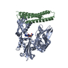
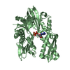
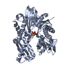
Entry Database : PDB / ID : 3ldoTitle Crystal structure of human GRP78 (70kDa heat shock protein 5 / BIP) ATPase domain in complex with AMPPNP 78 kDa glucose-regulated protein Keywords / / / / / / / / / / / / / / Function / homology Function Domain/homology Component
/ / / / / / / / / / / / / / / / / / / / / / / / / / / / / / / / / / / / / / / / / / / / / / / / / / / / / / / / / / / / / / / / / / / / / / / / / / / / / / / / / / / / / / / / / / / / / / / / / / / / / / / / / / Biological species Homo sapiens (human)Method / / / Resolution : 1.95 Å Authors Dokurno, P. / Surgenor, A.E. / Shaw, T. / Macias, A.T. / Massey, A.J. / Williamson, D.S. Journal : J.Med.Chem. / Year : 2011Title : Adenosine-Derived Inhibitors of 78 kDa Glucose Regulated Protein (Grp78) ATPase: Insights into Isoform Selectivity.Authors: Macias, A.T. / Williamson, D.S. / Allen, N. / Borgognoni, J. / Clay, A. / Daniels, Z. / Dokurno, P. / Drysdale, M.J. / Francis, G.L. / Graham, C.J. / Howes, R. / Matassova, N. / Murray, J.B. ... Authors : Macias, A.T. / Williamson, D.S. / Allen, N. / Borgognoni, J. / Clay, A. / Daniels, Z. / Dokurno, P. / Drysdale, M.J. / Francis, G.L. / Graham, C.J. / Howes, R. / Matassova, N. / Murray, J.B. / Parsons, R. / Shaw, T. / Surgenor, A.E. / Terry, L. / Wang, Y. / Wood, M. / Massey, A.J. History Deposition Jan 13, 2010 Deposition site / Processing site Revision 1.0 Jan 26, 2011 Provider / Type Revision 1.1 Jul 13, 2011 Group Revision 1.2 Feb 21, 2024 Group / Database references / Derived calculationsCategory chem_comp_atom / chem_comp_bond ... chem_comp_atom / chem_comp_bond / database_2 / struct_ref_seq_dif / struct_site Item _database_2.pdbx_DOI / _database_2.pdbx_database_accession ... _database_2.pdbx_DOI / _database_2.pdbx_database_accession / _struct_ref_seq_dif.details / _struct_site.pdbx_auth_asym_id / _struct_site.pdbx_auth_comp_id / _struct_site.pdbx_auth_seq_id
Show all Show less
 Movie
Movie Controller

 Yorodumi
 Open data
 Basic information
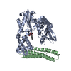
 Components
 Keywords
 Function and homology information
 Homo sapiens (human)
 X-RAY DIFFRACTION /
  Authors
 Citation
 Structure visualization
 Downloads & links
 Other downloads
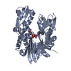


 PDBj
PDBj


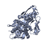
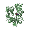
 Assembly
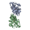
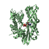


 Mass: 506.196 Da / Num. of mol.: 2 / Source method: obtained synthetically / Formula: C10H17N6O12P3 / Comment: AMP-PNP, energy-carrying molecule analogue*YM
 Mass: 18.015 Da / Num. of mol.: 499 / Source method: isolated from a natural source / Formula: H2O
 Sample preparation
 / Beamline: ID29 / Wavelength: 0.973 Å
 Processing