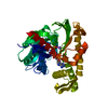
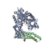
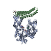
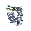
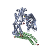
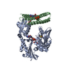
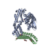
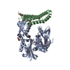
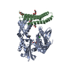
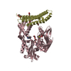
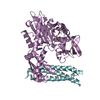
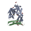
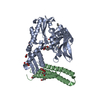
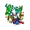
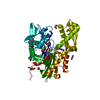
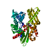
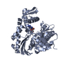
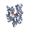
Entry Database : PDB / ID : 5ar0Title HSP72 with adenosine-derived inhibitor HEAT SHOCK 70 KDA PROTEIN 1A Keywords / / / / / / Function / homology Function Domain/homology Component
/ / / / / / / / / / / / / / / / / / / / / / / / / / / / / / / / / / / / / / / / / / / / / / / / / / / / / / / / / / / / / / / / / / / / / / / / / / / / / / / / / / / / / / / / / / / / / / / / / / / / / / / / / / / / / / / / / / / / / / / / / Biological species HOMO SAPIENS (human)Method / / / Resolution : 1.9 Å Authors Cheeseman, M.D. / Westwood, I.M. / Barbeau, O. / Rowlands, M.G. / Jones, A.M. / Jeganathan, F. / Burke, R. / Dobson, S.E. / Workman, P. / Collins, I. ...Cheeseman, M.D. / Westwood, I.M. / Barbeau, O. / Rowlands, M.G. / Jones, A.M. / Jeganathan, F. / Burke, R. / Dobson, S.E. / Workman, P. / Collins, I. / van Montfort, R.L.M. / Jones, K. Journal : J.Med.Chem. / Year : 2016Title : Exploiting Protein Conformational Change to Optimize Adenosine-Derived Inhibitors of Hsp70.Authors : Cheeseman, M.D. / Westwood, I.M. / Barbeau, O. / Rowlands, M.G. / Dobson, S. / Jones, A.M. / Jeganathan, F. / Burke, R. / Kadi, N. / Workman, P. / Collins, I. / Van Montfort, R.L.M. / Jones, K. History Deposition Sep 22, 2015 Deposition site / Processing site Revision 1.0 May 11, 2016 Provider / Type Revision 1.1 Jun 8, 2016 Group Revision 1.2 Jan 10, 2024 Group Data collection / Database references ... Data collection / Database references / Derived calculations / Other / Refinement description Category chem_comp_atom / chem_comp_bond ... chem_comp_atom / chem_comp_bond / database_2 / pdbx_database_status / pdbx_initial_refinement_model / struct_site Item _database_2.pdbx_DOI / _database_2.pdbx_database_accession ... _database_2.pdbx_DOI / _database_2.pdbx_database_accession / _pdbx_database_status.status_code_sf / _struct_site.pdbx_auth_asym_id / _struct_site.pdbx_auth_comp_id / _struct_site.pdbx_auth_seq_id
Show all Show less
 Movie
Movie Controller


 Open data
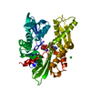
 Basic information
 Components
 Keywords
 Function and homology information
 HOMO SAPIENS (human)
 X-RAY DIFFRACTION /
  Authors
 Citation
 Structure visualization
 Downloads & links
 Other downloads
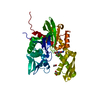


 PDBj
PDBj


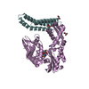
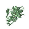
 Assembly
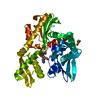

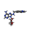


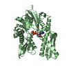
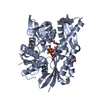
 Mass: 423.425 Da / Num. of mol.: 1 / Source method: obtained synthetically / Formula: C20H21N7O4
 Mass: 92.094 Da / Num. of mol.: 3 / Source method: obtained synthetically / Formula: C3H8O3
 Mass: 78.133 Da / Num. of mol.: 2 / Source method: obtained synthetically / Formula: C2H6OS / Comment: DMSO, precipitant*YM
 Mass: 35.453 Da / Num. of mol.: 1 / Source method: obtained synthetically / Formula: Cl
 Sample preparation
 / Beamline: I24 / Wavelength: 0.9686
  Processing