 Movie
Movie Controller

[English] 日本語
 Yorodumi
- PDB-5aqy: Fragment-based screening of HSP70 sheds light on the functional r... -
+ Open data

- Basic information
| Entry | Database: PDB / ID: 5aqy | ||||||
|---|---|---|---|---|---|---|---|
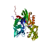
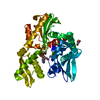
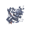
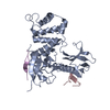
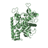
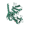
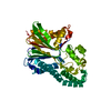
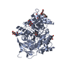
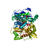
| Title | Fragment-based screening of HSP70 sheds light on the functional role of ATP-binding site residues | ||||||
 Components Components | HEAT SHOCK 70 KDA PROTEIN 1A | ||||||
 Keywords Keywords | CHAPERONE / HEAT SHOCK PROTEIN / HSP70 / HSP72 / HSC70 / ATPASE / BAG1 / FRAGMENT | ||||||
| Function / homology |  Function and homology information Function and homology information: / cellular heat acclimation / negative regulation of inclusion body assembly / Viral RNP Complexes in the Host Cell Nucleus / C3HC4-type RING finger domain binding / positive regulation of nucleotide-binding oligomerization domain containing 2 signaling pathway / positive regulation of microtubule nucleation / ATP-dependent protein disaggregase activity / misfolded protein binding / negative regulation of mitochondrial outer membrane permeabilization involved in apoptotic signaling pathway ...: / cellular heat acclimation / negative regulation of inclusion body assembly / Viral RNP Complexes in the Host Cell Nucleus / C3HC4-type RING finger domain binding / positive regulation of nucleotide-binding oligomerization domain containing 2 signaling pathway / positive regulation of microtubule nucleation / ATP-dependent protein disaggregase activity / misfolded protein binding / negative regulation of mitochondrial outer membrane permeabilization involved in apoptotic signaling pathway / regulation of mitotic spindle assembly / positive regulation of tumor necrosis factor-mediated signaling pathway / aggresome / lysosomal transport / cellular response to steroid hormone stimulus / mRNA catabolic process / regulation of protein ubiquitination / cellular response to unfolded protein / HSF1-dependent transactivation / Regulation of HSF1-mediated heat shock response / response to unfolded protein / Mitochondrial unfolded protein response (UPRmt) / negative regulation of extrinsic apoptotic signaling pathway in absence of ligand / Attenuation phase / chaperone-mediated protein complex assembly / negative regulation of endoplasmic reticulum stress-induced intrinsic apoptotic signaling pathway / transcription regulator inhibitor activity / ATP metabolic process / endoplasmic reticulum unfolded protein response / heat shock protein binding / protein folding chaperone / inclusion body / negative regulation of protein ubiquitination / positive regulation of erythrocyte differentiation / HSP90 chaperone cycle for steroid hormone receptors (SHR) in the presence of ligand / positive regulation of RNA splicing / : / positive regulation of interleukin-8 production / ATP-dependent protein folding chaperone / centriole / negative regulation of transforming growth factor beta receptor signaling pathway / AUF1 (hnRNP D0) binds and destabilizes mRNA / negative regulation of cell growth / PKR-mediated signaling / G protein-coupled receptor binding / histone deacetylase binding / disordered domain specific binding / : / transcription corepressor activity / positive regulation of proteasomal ubiquitin-dependent protein catabolic process / cellular response to heat / protein refolding / virus receptor activity / cellular response to oxidative stress / blood microparticle / vesicle / ficolin-1-rich granule lumen / nuclear speck / protein stabilization / cadherin binding / ribonucleoprotein complex / receptor ligand activity / signaling receptor binding / negative regulation of cell population proliferation / focal adhesion / Neutrophil degranulation / ubiquitin protein ligase binding / positive regulation of gene expression / centrosome / negative regulation of apoptotic process / perinuclear region of cytoplasm / enzyme binding / negative regulation of transcription by RNA polymerase II / endoplasmic reticulum / ATP hydrolysis activity / protein-containing complex / mitochondrion / : / RNA binding / extracellular exosome / extracellular region / nucleoplasm / ATP binding / nucleus / plasma membrane / cytoplasm / cytosol Similarity search - Function | ||||||
| Biological species |  HOMO SAPIENS (human) HOMO SAPIENS (human) | ||||||
| Method |  X-RAY DIFFRACTION / SYNCHROTRON / MOLECULAR REPLACEMENT / Resolution: 1.56 Å X-RAY DIFFRACTION / SYNCHROTRON / MOLECULAR REPLACEMENT / Resolution: 1.56 Å | ||||||
 Authors Authors | Jones, A.M. / Westwood, I.M. / Osborne, J.D. / Matthews, T.P. / Cheeseman, M.D. / Rowlands, M.G. / Jeganathan, F. / Burke, R. / Lee, D. / Kadi, N. ...Jones, A.M. / Westwood, I.M. / Osborne, J.D. / Matthews, T.P. / Cheeseman, M.D. / Rowlands, M.G. / Jeganathan, F. / Burke, R. / Lee, D. / Kadi, N. / Liu, M. / Richards, M. / McAndrew, C. / Yahya, N. / Dobson, S.E. / Jones, K. / Workman, P. / Collins, I. / van Montfort, R.L.M. | ||||||
 Citation Citation | Journal: Sci Rep / Year: 2016 Title: A fragment-based approach applied to a highly flexible target: Insights and challenges towards the inhibition of HSP70 isoforms. Authors: Jones, A.M. / Westwood, I.M. / Osborne, J.D. / Matthews, T.P. / Cheeseman, M.D. / Rowlands, M.G. / Jeganathan, F. / Burke, R. / Lee, D. / Kadi, N. / Liu, M. / Richards, M. / McAndrew, C. / ...Authors: Jones, A.M. / Westwood, I.M. / Osborne, J.D. / Matthews, T.P. / Cheeseman, M.D. / Rowlands, M.G. / Jeganathan, F. / Burke, R. / Lee, D. / Kadi, N. / Liu, M. / Richards, M. / McAndrew, C. / Yahya, N. / Dobson, S.E. / Jones, K. / Workman, P. / Collins, I. / van Montfort, R.L. | ||||||
| History |
|
- Structure visualization
| Structure viewer | Molecule: MolmilJmol/JSmol |
|---|
- Downloads & links
-Download
| PDBx/mmCIF format | 5aqy.cif.gz | 174 KB | Display | PDBx/mmCIF format |
|---|---|---|---|---|
| PDB format | pdb5aqy.ent.gz | 136.9 KB | Display | PDB format |
| PDBx/mmJSON format | 5aqy.json.gz | Tree view | PDBx/mmJSON format | |
| Others |  Other downloads Other downloads |
-Validation report
| Arichive directory | https://data.pdbj.org/pub/pdb/validation_reports/aq/5aqyftp://data.pdbj.org/pub/pdb/validation_reports/aq/5aqy | HTTPS FTP |
|---|
-Related structure data
| Related structure data |  5aqfC  5aqgC  5aqhC  5aqiC  5aqjC  5aqkC  5aqlC  5aqmC  5aqnC  5aqoC  5aqpC  5aqqC  5aqrC  5aqsC  5aqtC  5aquC  5aqvC 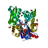 5aqwC  5aqxC 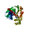 1s3xS C: citing same article ( S: Starting model for refinement |
|---|---|
| Similar structure data |


-Links
 PDBj
PDBj


- Assembly
| Deposited unit | 
| ||||||||
|---|---|---|---|---|---|---|---|---|---|
| 1 |
| ||||||||
| Unit cell |
|
-Components
-Protein , 1 types, 1 molecules A
| #1: Protein | Mass: 43195.902 Da / Num. of mol.: 1 / Fragment: NUCLEOTIDE BINDING DOMAIN, UNP RESIDUES 1-380 Source method: isolated from a genetically manipulated source Source: (gene. exp.) HOMO SAPIENS (human) / Production host:  |
|---|
-Non-polymers , 5 types, 345 molecules 


| #2: Chemical | ChemComp-ADN /  Mass: 267.241 Da / Num. of mol.: 1 / Source method: obtained synthetically / Formula: C10H13N5O4 Mass: 267.241 Da / Num. of mol.: 1 / Source method: obtained synthetically / Formula: C10H13N5O4 | ||||||
|---|---|---|---|---|---|---|---|
| #3: Chemical | ChemComp-EDO /  Mass: 62.068 Da / Num. of mol.: 21 / Source method: obtained synthetically / Formula: C2H6O2 Mass: 62.068 Da / Num. of mol.: 21 / Source method: obtained synthetically / Formula: C2H6O2#4: Chemical | ChemComp-DMS / |  Mass: 78.133 Da / Num. of mol.: 1 / Source method: obtained synthetically / Formula: C2H6OS / Comment: DMSO, precipitant*YM Mass: 78.133 Da / Num. of mol.: 1 / Source method: obtained synthetically / Formula: C2H6OS / Comment: DMSO, precipitant*YM#5: Chemical | ChemComp-MG / |  Mass: 24.305 Da / Num. of mol.: 1 / Source method: obtained synthetically / Formula: Mg Mass: 24.305 Da / Num. of mol.: 1 / Source method: obtained synthetically / Formula: Mg#6: Water | ChemComp-HOH / | Mass: 18.015 Da / Num. of mol.: 321 / Source method: isolated from a natural source / Formula: H2O |
-Details
| Sequence details | RESIDUES 382 ONWARDS ARE A CLONING ARTEFACT DERIVED FROM THE EXPRESSION |
|---|
-Experimental details
-Experiment
| Experiment | Method: X-RAY DIFFRACTION / Number of used crystals: 1 |
|---|
- Sample preparation
| Crystal | Density Matthews: 2.4 Å3/Da / Density % sol: 48.8 % / Description: NONE |
|---|---|
| Crystal grow | pH: 7.5 Details: 17-28% (W/V) PEG3350, 0.1M HEPES PH 7.5, 2MM MGCL2, 2MM NAH2PO4 AND 5MM ADENOSINE |
-Data collection
| Diffraction | Mean temperature: 100 K |
|---|---|
| Diffraction source | Source: SYNCHROTRON / Site: Diamond  / Beamline: I02 / Wavelength: 0.9728 / Beamline: I02 / Wavelength: 0.9728 |
| Detector | Type: ADSC QUANTUM 315 / Detector: CCD / Date: Dec 17, 2008 |
| Radiation | Protocol: SINGLE WAVELENGTH / Monochromatic (M) / Laue (L): M / Scattering type: x-ray |
| Radiation wavelength | Wavelength: 0.9728 Å / Relative weight: 1 |
| Reflection | Resolution: 1.56→47.97 Å / Num. obs: 58145 / % possible obs: 98.9 % / Observed criterion σ(I): 0 / Redundancy: 4.9 % / Biso Wilson estimate: 20.9 Å2 / Rmerge(I) obs: 0.08 / Net I/σ(I): 5.2 |
| Reflection shell | Resolution: 1.56→1.59 Å / Redundancy: 3.7 % / Rmerge(I) obs: 1.3 / Mean I/σ(I) obs: 1 / % possible all: 97 |
- Processing
| Software |
| ||||||||||||||||||||||||||||||||||||||||||||||||||||||||||||||||||||||||||||||||||||||||||||||||||||||||||||||||||||||||||||||||||||||||||||||||||||||||||||||||||||||||||||||||||||||||||||||||||||||||||||||||||||||||||||||||||||||||||||||||||||||||||||||||||||||||||||||||||||||||||||||||||||||||||||
|---|---|---|---|---|---|---|---|---|---|---|---|---|---|---|---|---|---|---|---|---|---|---|---|---|---|---|---|---|---|---|---|---|---|---|---|---|---|---|---|---|---|---|---|---|---|---|---|---|---|---|---|---|---|---|---|---|---|---|---|---|---|---|---|---|---|---|---|---|---|---|---|---|---|---|---|---|---|---|---|---|---|---|---|---|---|---|---|---|---|---|---|---|---|---|---|---|---|---|---|---|---|---|---|---|---|---|---|---|---|---|---|---|---|---|---|---|---|---|---|---|---|---|---|---|---|---|---|---|---|---|---|---|---|---|---|---|---|---|---|---|---|---|---|---|---|---|---|---|---|---|---|---|---|---|---|---|---|---|---|---|---|---|---|---|---|---|---|---|---|---|---|---|---|---|---|---|---|---|---|---|---|---|---|---|---|---|---|---|---|---|---|---|---|---|---|---|---|---|---|---|---|---|---|---|---|---|---|---|---|---|---|---|---|---|---|---|---|---|---|---|---|---|---|---|---|---|---|---|---|---|---|---|---|---|---|---|---|---|---|---|---|---|---|---|---|---|---|---|---|---|---|---|---|---|---|---|---|---|---|---|---|---|---|---|---|---|---|---|---|---|---|---|---|---|---|---|---|---|---|---|---|---|---|---|---|---|---|---|---|---|---|---|---|---|---|---|---|---|---|---|---|
| Refinement | Method to determine structure: MOLECULAR REPLACEMENT Starting model: PDB ENTRY 1S3X Resolution: 1.56→47.97 Å / Cor.coef. Fo:Fc: 0.9582 / Cor.coef. Fo:Fc free: 0.9477 / SU R Cruickshank DPI: 0.076 / Cross valid method: THROUGHOUT / σ(F): 0 / SU R Blow DPI: 0.079 / SU Rfree Blow DPI: 0.08 / SU Rfree Cruickshank DPI: 0.077
| ||||||||||||||||||||||||||||||||||||||||||||||||||||||||||||||||||||||||||||||||||||||||||||||||||||||||||||||||||||||||||||||||||||||||||||||||||||||||||||||||||||||||||||||||||||||||||||||||||||||||||||||||||||||||||||||||||||||||||||||||||||||||||||||||||||||||||||||||||||||||||||||||||||||||||||
| Displacement parameters | Biso mean: 28.08 Å2
| ||||||||||||||||||||||||||||||||||||||||||||||||||||||||||||||||||||||||||||||||||||||||||||||||||||||||||||||||||||||||||||||||||||||||||||||||||||||||||||||||||||||||||||||||||||||||||||||||||||||||||||||||||||||||||||||||||||||||||||||||||||||||||||||||||||||||||||||||||||||||||||||||||||||||||||
| Refine analyze | Luzzati coordinate error obs: 0.205 Å | ||||||||||||||||||||||||||||||||||||||||||||||||||||||||||||||||||||||||||||||||||||||||||||||||||||||||||||||||||||||||||||||||||||||||||||||||||||||||||||||||||||||||||||||||||||||||||||||||||||||||||||||||||||||||||||||||||||||||||||||||||||||||||||||||||||||||||||||||||||||||||||||||||||||||||||
| Refinement step | Cycle: LAST / Resolution: 1.56→47.97 Å
| ||||||||||||||||||||||||||||||||||||||||||||||||||||||||||||||||||||||||||||||||||||||||||||||||||||||||||||||||||||||||||||||||||||||||||||||||||||||||||||||||||||||||||||||||||||||||||||||||||||||||||||||||||||||||||||||||||||||||||||||||||||||||||||||||||||||||||||||||||||||||||||||||||||||||||||
| Refine LS restraints |
| ||||||||||||||||||||||||||||||||||||||||||||||||||||||||||||||||||||||||||||||||||||||||||||||||||||||||||||||||||||||||||||||||||||||||||||||||||||||||||||||||||||||||||||||||||||||||||||||||||||||||||||||||||||||||||||||||||||||||||||||||||||||||||||||||||||||||||||||||||||||||||||||||||||||||||||
| LS refinement shell | Resolution: 1.56→1.6 Å / Total num. of bins used: 20
| ||||||||||||||||||||||||||||||||||||||||||||||||||||||||||||||||||||||||||||||||||||||||||||||||||||||||||||||||||||||||||||||||||||||||||||||||||||||||||||||||||||||||||||||||||||||||||||||||||||||||||||||||||||||||||||||||||||||||||||||||||||||||||||||||||||||||||||||||||||||||||||||||||||||||||||
| Refinement TLS params. | Method: refined / Refine-ID: X-RAY DIFFRACTION
|
