 Movie
Movie Controller

[English] 日本語
 Yorodumi
- PDB-2h9d: Pyruvate-Bound Structure of the Isochorismate-Pyruvate Lyase from... -
+ Open data

- Basic information
| Entry | Database: PDB / ID: 2h9d | |||||||||
|---|---|---|---|---|---|---|---|---|---|---|
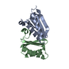
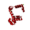
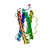
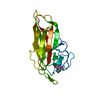
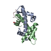
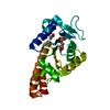
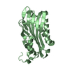
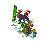
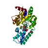
| Title | Pyruvate-Bound Structure of the Isochorismate-Pyruvate Lyase from Pseudomonas aerugionsa | |||||||||
 Components Components | Salicylate biosynthesis protein pchB | |||||||||
 Keywords Keywords | LYASE / intertwined dimer | |||||||||
| Function / homology |  Function and homology information Function and homology informationpyochelin biosynthetic process / isochorismate lyase / carbon-oxygen lyase activity / isochorismate pyruvate lyase activity / salicylic acid biosynthetic process / chorismate metabolic process / chorismate mutase / chorismate mutase activity Similarity search - Function | |||||||||
| Biological species |   Pseudomonas aeruginosa (bacteria) Pseudomonas aeruginosa (bacteria) | |||||||||
| Method |  X-RAY DIFFRACTION / MOLECULAR REPLACEMENT / Resolution: 1.95 Å X-RAY DIFFRACTION / MOLECULAR REPLACEMENT / Resolution: 1.95 Å | |||||||||
 Authors Authors | Lamb, A.L. / Zaitseva, J. / Lu, J. | |||||||||
 Citation Citation | Journal: J.Biol.Chem. / Year: 2006 Title: Two Crystal Structures of the Isochorismate Pyruvate Lyase from Pseudomonas aeruginosa. Authors: Zaitseva, J. / Lu, J. / Olechoski, K.L. / Lamb, A.L. | |||||||||
| History |
|
- Structure visualization
| Structure viewer | Molecule: MolmilJmol/JSmol |
|---|
- Downloads & links
-Download
| PDBx/mmCIF format | 2h9d.cif.gz | 90.3 KB | Display | PDBx/mmCIF format |
|---|---|---|---|---|
| PDB format | pdb2h9d.ent.gz | 68.6 KB | Display | PDB format |
| PDBx/mmJSON format | 2h9d.json.gz | Tree view | PDBx/mmJSON format | |
| Others |  Other downloads Other downloads |
-Validation report
| Arichive directory | https://data.pdbj.org/pub/pdb/validation_reports/h9/2h9dftp://data.pdbj.org/pub/pdb/validation_reports/h9/2h9d | HTTPS FTP |
|---|
-Related structure data
| Related structure data | 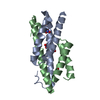 2h9cSC S: Starting model for refinement C: citing same article ( |
|---|---|
| Similar structure data |

-Links
 PDBj
PDBj


- Assembly
| Deposited unit | 
| ||||||||
|---|---|---|---|---|---|---|---|---|---|
| 1 | 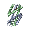
| ||||||||
| 2 | 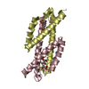
| ||||||||
| 3 |
| ||||||||
| 4 | 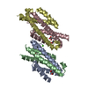
| ||||||||
| Unit cell |
| ||||||||
| Details | The asymmetric unit contains 2 dimers. Each dimer respresents one biological assembly. |
-Components
| #1: Protein | Mass: 11447.064 Da / Num. of mol.: 4 Source method: isolated from a genetically manipulated source Source: (gene. exp.) Pseudomonas aeruginosa (bacteria) / Strain: PAO1 / Gene: PchB / Plasmid: pET29b / Species (production host): Escherichia coli / Production host: References: UniProt: Q51507, Lyases; Carbon-carbon lyases; Other carbon-carbon lyases #2: Chemical | ChemComp-PYR /   Mass: 88.062 Da / Num. of mol.: 6 / Source method: obtained synthetically / Formula: C3H4O3 Mass: 88.062 Da / Num. of mol.: 6 / Source method: obtained synthetically / Formula: C3H4O3#3: Chemical | ChemComp-CA / |   Mass: 40.078 Da / Num. of mol.: 1 / Source method: obtained synthetically / Formula: Ca Mass: 40.078 Da / Num. of mol.: 1 / Source method: obtained synthetically / Formula: Ca#4: Water | ChemComp-HOH / |  Mass: 18.015 Da / Num. of mol.: 181 / Source method: isolated from a natural source / Formula: H2O Mass: 18.015 Da / Num. of mol.: 181 / Source method: isolated from a natural source / Formula: H2O |
|---|
-Experimental details
-Experiment
| Experiment | Method: X-RAY DIFFRACTION / Number of used crystals: 1 |
|---|
- Sample preparation
| Crystal | Density Matthews: 1.97 Å3/Da / Density % sol: 37.7 % |
|---|---|
| Crystal grow | Temperature: 298 K / Method: vapor diffusion, hanging drop / pH: 7 Details: 100 mM ADA, 25% PEG 3350, 0.15 M calcium acetate, 10% glycerol, pH 7.0, VAPOR DIFFUSION, HANGING DROP, temperature 298K |
-Data collection
| Diffraction | Mean temperature: 138 K |
|---|---|
| Diffraction source | Source: ROTATING ANODE / Type: RIGAKU RU200 / Wavelength: 1.5418 Å |
| Detector | Type: RIGAKU RAXIS IV / Detector: IMAGE PLATE / Date: Sep 22, 2005 |
| Radiation | Monochromator: osmic mirrors / Protocol: SINGLE WAVELENGTH / Monochromatic (M) / Laue (L): M / Scattering type: x-ray |
| Radiation wavelength | Wavelength: 1.5418 Å / Relative weight: 1 |
| Reflection | Resolution: 1.95→57.2 Å / Num. obs: 25853 / % possible obs: 95 % / Observed criterion σ(F): 1 / Observed criterion σ(I): 1 / Biso Wilson estimate: 26.8 Å2 / Rmerge(I) obs: 0.066 |
| Reflection shell | Resolution: 1.95→2.02 Å / Rmerge(I) obs: 0.405 / % possible all: 75.7 |
- Processing
| Software |
| |||||||||||||||||||||||||
|---|---|---|---|---|---|---|---|---|---|---|---|---|---|---|---|---|---|---|---|---|---|---|---|---|---|---|
| Refinement | Method to determine structure: MOLECULAR REPLACEMENT Starting model: PDB ENTRY 2H9C Resolution: 1.95→57.2 Å / σ(F): 0 / Stereochemistry target values: Engh & Huber
| |||||||||||||||||||||||||
| Displacement parameters | Biso mean: 28.6 Å2 | |||||||||||||||||||||||||
| Refinement step | Cycle: LAST / Resolution: 1.95→57.2 Å
| |||||||||||||||||||||||||
| Refine LS restraints |
|
