 Movie
Movie Controller

[English] 日本語
 Yorodumi
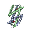
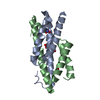
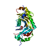
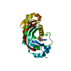
- PDB-3g1c: The crystal structure of a TrpR like protein from Eubacterium eli... -
+ Open data

- Basic information
| Entry | Database: PDB / ID: 3g1c | ||||||
|---|---|---|---|---|---|---|---|
| Title | The crystal structure of a TrpR like protein from Eubacterium eligens ATCC 27750 | ||||||
 Components Components | The TrpR like protein from Eubacterium eligens ATCC 27750 | ||||||
 Keywords Keywords | transcription / DNA binding protein / TrpR like protein / Eubacterium eligens / Structural Genomics / PSI / MCSG / Protein Structure Initiative / Midwest Center for Structural Genomics / unknown function | ||||||
| Function / homology |  Function and homology information Function and homology informationsequence-specific DNA binding / DNA-binding transcription factor activity Similarity search - Function | ||||||
| Biological species |  Eubacterium eligens (bacteria) Eubacterium eligens (bacteria) | ||||||
| Method |  X-RAY DIFFRACTION / SYNCHROTRON / SAD / Resolution: 2.2 Å X-RAY DIFFRACTION / SYNCHROTRON / SAD / Resolution: 2.2 Å | ||||||
 Authors Authors | Zhang, R. / Hendricks, R. / Freeman, L. / Babnigg, G. / Joachimiak, A. / Midwest Center for Structural Genomics (MCSG) | ||||||
 Citation Citation | Journal: To be Published Title: The crystal structure of a TrpR like protein from Eubacterium eligens ATCC 27750 Authors: Zhang, R. / Hendricks, R. / Freeman, L. / Babnigg, G. / Joachimiak, A. | ||||||
| History |
|
- Structure visualization
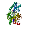
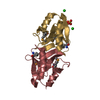
| Structure viewer | Molecule: MolmilJmol/JSmol |
|---|
- Downloads & links
-Download
| PDBx/mmCIF format | 3g1c.cif.gz | 32.4 KB | Display | PDBx/mmCIF format |
|---|---|---|---|---|
| PDB format | pdb3g1c.ent.gz | 22.2 KB | Display | PDB format |
| PDBx/mmJSON format | 3g1c.json.gz | Tree view | PDBx/mmJSON format | |
| Others |  Other downloads Other downloads |
-Validation report
| Arichive directory | https://data.pdbj.org/pub/pdb/validation_reports/g1/3g1cftp://data.pdbj.org/pub/pdb/validation_reports/g1/3g1c | HTTPS FTP |
|---|
-Related structure data
| Similar structure data | |
|---|---|
| Other databases |


-Links
 PDBj
PDBj
- Assembly
| Deposited unit | 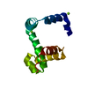
| ||||||||
|---|---|---|---|---|---|---|---|---|---|
| 1 | 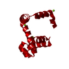
| ||||||||
| Unit cell |
| ||||||||
| Details | The second part of the biological assembly is generated by the two fold axis: -y,-x,-z+1/2 |
-Components
| #1: Protein | Mass: 12059.517 Da / Num. of mol.: 1 Source method: isolated from a genetically manipulated source Source: (gene. exp.) Eubacterium eligens (bacteria) / Strain: ATCC 27750 / Plasmid: pMCSG19 / Production host: | ||
|---|---|---|---|
| #2: Chemical |   Mass: 24.305 Da / Num. of mol.: 3 / Source method: obtained synthetically / Formula: Mg Mass: 24.305 Da / Num. of mol.: 3 / Source method: obtained synthetically / Formula: Mg#3: Water | ChemComp-HOH / |  Mass: 18.015 Da / Num. of mol.: 59 / Source method: isolated from a natural source / Formula: H2O Mass: 18.015 Da / Num. of mol.: 59 / Source method: isolated from a natural source / Formula: H2O |
-Experimental details
-Experiment
| Experiment | Method: X-RAY DIFFRACTION / Number of used crystals: 1 |
|---|
- Sample preparation
| Crystal | Density Matthews: 3.12 Å3/Da / Density % sol: 60.58 % |
|---|---|
| Crystal grow | Temperature: 289 K / Method: vapor diffusion, sitting drop / pH: 7.5 Details: 0.1M HEPES pH7.5, 0.5M Magnesium formate dihydrate, VAPOR DIFFUSION, SITTING DROP, temperature 289K |
-Data collection
| Diffraction | Mean temperature: 100 K |
|---|---|
| Diffraction source | Source: SYNCHROTRON / Site: APS  / Beamline: 19-ID / Wavelength: 0.9794 Å / Beamline: 19-ID / Wavelength: 0.9794 Å |
| Detector | Type: ADSC QUANTUM 315 / Detector: CCD / Date: Jul 3, 2008 / Details: mirrors |
| Radiation | Monochromator: Si 111 channel / Protocol: SINGLE WAVELENGTH / Monochromatic (M) / Laue (L): M / Scattering type: x-ray |
| Radiation wavelength | Wavelength: 0.9794 Å / Relative weight: 1 |
| Reflection | Resolution: 2.2→38.63 Å / Num. all: 7945 / Num. obs: 7885 / % possible obs: 99.24 % / Observed criterion σ(F): 2 / Observed criterion σ(I): 2 / Redundancy: 15.4 % / Biso Wilson estimate: 41 Å2 / Rmerge(I) obs: 0.106 / Net I/σ(I): 31.34 |
| Reflection shell | Resolution: 2.2→2.257 Å / Redundancy: 10.8 % / Rmerge(I) obs: 0.548 / Mean I/σ(I) obs: 1.28 / Num. unique all: 594 / % possible all: 94.44 |
- Processing
| Software |
| |||||||||||||||||||||||||||||||||||||||||||||||||||||||||||||||||||||||||||||||||||||
|---|---|---|---|---|---|---|---|---|---|---|---|---|---|---|---|---|---|---|---|---|---|---|---|---|---|---|---|---|---|---|---|---|---|---|---|---|---|---|---|---|---|---|---|---|---|---|---|---|---|---|---|---|---|---|---|---|---|---|---|---|---|---|---|---|---|---|---|---|---|---|---|---|---|---|---|---|---|---|---|---|---|---|---|---|---|---|
| Refinement | Method to determine structure: SAD / Resolution: 2.2→38.63 Å / Cor.coef. Fo:Fc: 0.954 / Cor.coef. Fo:Fc free: 0.944 / SU B: 5.544 / SU ML: 0.135 / Cross valid method: THROUGHOUT / σ(F): 0 / σ(I): 1.28 / ESU R: 0.2 / ESU R Free: 0.174 Stereochemistry target values: MAXIMUM LIKELIHOOD WITH PHASES Details: HYDROGENS HAVE BEEN ADDED IN THE RIDING POSITIONS
| |||||||||||||||||||||||||||||||||||||||||||||||||||||||||||||||||||||||||||||||||||||
| Solvent computation | Ion probe radii: 0.8 Å / Shrinkage radii: 0.8 Å / VDW probe radii: 1.2 Å / Solvent model: MASK | |||||||||||||||||||||||||||||||||||||||||||||||||||||||||||||||||||||||||||||||||||||
| Displacement parameters | Biso mean: 39.638 Å2
| |||||||||||||||||||||||||||||||||||||||||||||||||||||||||||||||||||||||||||||||||||||
| Refinement step | Cycle: LAST / Resolution: 2.2→38.63 Å
| |||||||||||||||||||||||||||||||||||||||||||||||||||||||||||||||||||||||||||||||||||||
| Refine LS restraints |
| |||||||||||||||||||||||||||||||||||||||||||||||||||||||||||||||||||||||||||||||||||||
| LS refinement shell | Resolution: 2.2→2.257 Å / Total num. of bins used: 20
|
