Mass: 18.015 Da / Num. of mol.: 185 / Source method: isolated from a natural source / Formula: H2O
Has protein modification
Y
Sequence details
THE CONSTRUCT (1-216) WAS EXPRESSED WITH A PURIFICATION TAG MGSDKIHHHHHHENLYFQG. THE TAG WAS ...THE CONSTRUCT (1-216) WAS EXPRESSED WITH A PURIFICATION TAG MGSDKIHHHHHHENLYFQG. THE TAG WAS REMOVED WITH TEV PROTEASE LEAVING ONLY A GLYCINE (0) FOLLOWED BY THE TARGET SEQUENCE.
-
Experimental details
-
Experiment
Experiment
Method: X-RAY DIFFRACTION / Number of used crystals: 1
-
Sample preparation
Crystal
Density Matthews: 2.33 Å3/Da / Density % sol: 47.23 %
Crystal grow
Temperature: 277 K / Method: vapor diffusion, sitting drop Details: 14.90% polyethylene glycol 3350 0.3570M magnesium chloride, NANODROP, VAPOR DIFFUSION, SITTING DROP, temperature 277K
Type: ADSC QUANTUM 315 / Detector: CCD / Date: Apr 19, 2008 Details: 2-crystal monochromator, Si111, 1m long Rh coated bent cylindrical mirror for horizontal and vertical focussing
Radiation
Monochromator: 2-crystal, Si111 / Protocol: SINGLE WAVELENGTH / Monochromatic (M) / Laue (L): M / Scattering type: x-ray
Radiation wavelength
Wavelength: 0.978662 Å / Relative weight: 1
Reflection
Resolution: 2.15→39.958 Å / Num. obs: 25747 / % possible obs: 99.7 % / Observed criterion σ(I): -3 / Redundancy: 4.71 % / Biso Wilson estimate: 30.743 Å2 / Rmerge(I) obs: 0.119 / Net I/σ(I): 9.36
Reflection shell
Diffraction-ID: 1
Resolution (Å)
Redundancy (%)
Rmerge(I) obs
Mean I/σ(I) obs
Num. measured obs
Num. unique obs
% possible all
2.15-2.23
4.76
0.88
1.95
12336
2594
99.5
2.23-2.32
0.678
2.5
12031
2519
99.7
2.32-2.42
0.489
3.3
11597
2403
99.8
2.42-2.55
0.377
4.1
12531
2594
99.7
2.55-2.71
0.295
5
12155
2526
100
2.71-2.92
0.215
6.8
12389
2576
99.9
2.92-3.21
0.129
10.2
12046
2523
100
3.21-3.67
0.074
15.8
12088
2584
99.8
3.67-4.61
0.053
20.6
11944
2604
99.5
4.61-39.96
0.057
21.5
12064
2824
99.3
-
Phasing
Phasing
Method: SAD
-
Processing
Software
Name
Version
Classification
NB
MolProbity
3beta29
modelbuilding
PDB_EXTRACT
3.1
dataextraction
SHELX
phasing
SHARP
phasing
XSCALE
January10, 2014BUILT=20140307
datascaling
BUSTER-TNT
2.10.0
refinement
XDS
datareduction
SHELXD
phasing
BUSTER
2.10.0
refinement
Refinement
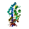
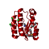
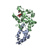
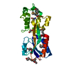
Method to determine structure: SAD / Resolution: 2.15→39.958 Å / Cor.coef. Fo:Fc: 0.933 / Cor.coef. Fo:Fc free: 0.9234 / Occupancy max: 1 / Occupancy min: 0.33 / Cross valid method: THROUGHOUT / σ(F): 0 Details: 1. A MET-INHIBITION PROTOCOL WAS USED FOR SELENOMETHIONINE INCORPORATION DURING PROTEIN EXPRESSION. THE OCCUPANCY OF THE SE ATOMS IN THE MSE RESIDUES WAS REDUCED TO 0.75 FOR THE REDUCED ...Details: 1. A MET-INHIBITION PROTOCOL WAS USED FOR SELENOMETHIONINE INCORPORATION DURING PROTEIN EXPRESSION. THE OCCUPANCY OF THE SE ATOMS IN THE MSE RESIDUES WAS REDUCED TO 0.75 FOR THE REDUCED SCATTERING POWER DUE TO PARTIAL S-MET INCORPORATION. 2. ATOM RECORD CONTAINS SUM OF TLS AND RESIDUAL B FACTORS. ANISOU RECORD CONTAINS SUM OF TLS AND RESIDUAL U FACTORS. 3. THE SAD PHASES WERE USED AS RESTRAINTS DURING REFINEMENT. 4. NCS RESTRAINTS WERE APPLIED USING BUSTER'S LSSR RESTRAINT REPRESENTATION (-AUTONCS). 5.MAGNESIUM (MG) FROM THE CRYSTALLIZATION SOLUTION AND 1,2-ETHANEDIOL (EDO) USED AS A CRYOPROTECTANT WERE MODELED INTO THE STRUCTURE. 6. THERE IS GAP IN THE MODEL ON SUBUNIT B BETWEEN RESIDUES 62-71 SINCE ELECTRON DENSITIES FOR THESE RESIDUES ARE DISORDERED.
In the structure databanks used in Yorodumi, some data are registered as the other names, "COVID-19 virus" and "2019-nCoV". Here are the details of the virus and the list of structure data.
Jan 31, 2019. EMDB accession codes are about to change! (news from PDBe EMDB page)
EMDB accession codes are about to change! (news from PDBe EMDB page)
The allocation of 4 digits for EMDB accession codes will soon come to an end. Whilst these codes will remain in use, new EMDB accession codes will include an additional digit and will expand incrementally as the available range of codes is exhausted. The current 4-digit format prefixed with “EMD-” (i.e. EMD-XXXX) will advance to a 5-digit format (i.e. EMD-XXXXX), and so on. It is currently estimated that the 4-digit codes will be depleted around Spring 2019, at which point the 5-digit format will come into force.
The EM Navigator/Yorodumi systems omit the EMD- prefix.
Related info.:Q: What is EMD? / ID/Accession-code notation in Yorodumi/EM Navigator
Yorodumi is a browser for structure data from EMDB, PDB, SASBDB, etc.
This page is also the successor to EM Navigator detail page, and also detail information page/front-end page for Omokage search.
The word "yorodu" (or yorozu) is an old Japanese word meaning "ten thousand". "mi" (miru) is to see.
Related info.:EMDB / PDB / SASBDB / Comparison of 3 databanks / Yorodumi Search / Aug 31, 2016. New EM Navigator & Yorodumi / Yorodumi Papers / Jmol/JSmol / Function and homology information / Changes in new EM Navigator and Yorodumi
 Movie
Movie Controller

 Yorodumi
 Open data
 Basic information
 Components
 Keywords
 Function and homology information
 Geobacter sulfurreducens PCA (bacteria)
 X-RAY DIFFRACTION /
  Authors
 Citation
 Structure visualization
 Downloads & links
 Other downloads


 PDBj
PDBj
 Assembly


 Mass: 24.305 Da / Num. of mol.: 2 / Source method: obtained synthetically / Formula: Mg
 Mass: 62.068 Da / Num. of mol.: 5 / Source method: obtained synthetically / Formula: C2H6O2
 Mass: 18.015 Da / Num. of mol.: 185 / Source method: isolated from a natural source / Formula: H2O
 Sample preparation
 / Beamline: BL1-5 / Wavelength: 0.978662
  Processing