 Movie
Movie Controller

[English] 日本語
 Yorodumi
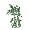
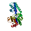
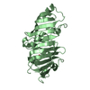
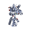
- PDB-1lvh: The Structure of Phosphorylated beta-phosphoglucomutase from Lact... -
+ Open data

- Basic information
| Entry | Database: PDB / ID: 1lvh | ||||||
|---|---|---|---|---|---|---|---|
| Title | The Structure of Phosphorylated beta-phosphoglucomutase from Lactoccocus lactis to 2.3 angstrom resolution | ||||||
 Components Components | beta-phosphoglucomutase | ||||||
 Keywords Keywords | ISOMERASE / HAD superfamily / phosphoaspartate / aspartylphosphate | ||||||
| Function / homology |  Function and homology information Function and homology informationbeta-phosphoglucomutase / beta-phosphoglucomutase activity / carbohydrate metabolic process / magnesium ion binding / cytoplasm Similarity search - Function | ||||||
| Biological species |  Lactococcus lactis (lactic acid bacteria) Lactococcus lactis (lactic acid bacteria) | ||||||
| Method |  X-RAY DIFFRACTION / SYNCHROTRON / MAD / Resolution: 2.3 Å X-RAY DIFFRACTION / SYNCHROTRON / MAD / Resolution: 2.3 Å | ||||||
 Authors Authors | Lahiri, S.D. / Zhang, G. / Dunaway-Mariano, D. / Allen, K.N. | ||||||
 Citation Citation | Journal: Biochemistry / Year: 2002 Title: Caught in the act: the structure of phosphorylated beta-phosphoglucomutase from Lactococcus lactis. Authors: Lahiri, S.D. / Zhang, G. / Dunaway-Mariano, D. / Allen, K.N. #1: Journal: Acta Crystallogr.,Sect.D / Year: 2002Title: Crystallization and preliminary X-ray diffraction studies of beta-phosphoglucomutase from Lactococcus lactus Authors: Lahiri, S.D. / Zhang, G. / Radstrom, P. / Dunaway-Mariano, D. / Allen, K.N. | ||||||
| History |
|
- Structure visualization
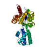
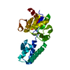
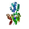
| Structure viewer | Molecule: MolmilJmol/JSmol |
|---|
- Downloads & links
-Download
| PDBx/mmCIF format | 1lvh.cif.gz | 102.8 KB | Display | PDBx/mmCIF format |
|---|---|---|---|---|
| PDB format | pdb1lvh.ent.gz | 79.2 KB | Display | PDB format |
| PDBx/mmJSON format | 1lvh.json.gz | Tree view | PDBx/mmJSON format | |
| Others |  Other downloads Other downloads |
-Validation report
| Arichive directory | https://data.pdbj.org/pub/pdb/validation_reports/lv/1lvhftp://data.pdbj.org/pub/pdb/validation_reports/lv/1lvh | HTTPS FTP |
|---|
-Related structure data
| Related structure data | |
|---|---|
| Similar structure data |


-Links
 PDBj
PDBj

- Assembly
| Deposited unit | 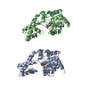
| ||||||||
|---|---|---|---|---|---|---|---|---|---|
| 1 | 
| ||||||||
| 2 | 
| ||||||||
| Unit cell |
| ||||||||
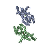
| Details | The biological assembly is a monomer |
-Components
| #1: Protein | Mass: 24319.574 Da / Num. of mol.: 2 Source method: isolated from a genetically manipulated source Source: (gene. exp.) Lactococcus lactis (lactic acid bacteria)Gene: PGMB / Plasmid: pET3A / Species (production host): Escherichia coli / Production host: #2: Chemical |   Mass: 24.305 Da / Num. of mol.: 2 / Source method: obtained synthetically / Formula: Mg Mass: 24.305 Da / Num. of mol.: 2 / Source method: obtained synthetically / Formula: Mg#3: Water | ChemComp-HOH / |  Mass: 18.015 Da / Num. of mol.: 259 / Source method: isolated from a natural source / Formula: H2O Mass: 18.015 Da / Num. of mol.: 259 / Source method: isolated from a natural source / Formula: H2OHas protein modification | Y | |
|---|
-Experimental details
-Experiment
| Experiment | Method: X-RAY DIFFRACTION / Number of used crystals: 1 |
|---|
- Sample preparation
| Crystal | Density Matthews: 2.85 Å3/Da / Density % sol: 56.91 % | ||||||||||||||||||||||||
|---|---|---|---|---|---|---|---|---|---|---|---|---|---|---|---|---|---|---|---|---|---|---|---|---|---|
| Crystal grow | Temperature: 291 K / Method: vapor diffusion, hanging drop / pH: 6.1 Details: 16% PEG 3350, 0.15M Ammonium Floride, pH 6.1, VAPOR DIFFUSION, HANGING DROP, temperature 291K | ||||||||||||||||||||||||
| Crystal grow | *PLUS pH: 6.5 Details: Lahiri, S.D., (2002) Acta Crystallogr., Sect.D, 58, 324. | ||||||||||||||||||||||||
| Components of the solutions | *PLUS
|
-Data collection
| Diffraction | Mean temperature: 93 K | ||||||||||||
|---|---|---|---|---|---|---|---|---|---|---|---|---|---|
| Diffraction source | Source: SYNCHROTRON / Site: APS  / Beamline: 14-BM-D / Wavelength: 0.9792369, 0.9790073, 0.96112713 / Beamline: 14-BM-D / Wavelength: 0.9792369, 0.9790073, 0.96112713 | ||||||||||||
| Detector | Type: ADSC QUANTUM 4 / Detector: CCD / Date: Mar 3, 2001 / Details: Bent cylindrical Si-mirror (Rh coating) | ||||||||||||
| Radiation | Monochromator: Si(111) double-crystal monochromator / Protocol: MAD / Monochromatic (M) / Laue (L): M / Scattering type: x-ray | ||||||||||||
| Radiation wavelength |
| ||||||||||||
| Reflection | Resolution: 2.3→50 Å / Num. all: 25469 / Num. obs: 25469 / % possible obs: 91 % / Observed criterion σ(F): 0 / Observed criterion σ(I): 0 / Redundancy: 16.5 % / Biso Wilson estimate: 19.1 Å2 / Rmerge(I) obs: 0.081 / Rsym value: 0.081 / Net I/σ(I): 12.3 | ||||||||||||
| Reflection shell | Resolution: 2.3→2.38 Å / Redundancy: 3.5 % / Rmerge(I) obs: 0.191 / Mean I/σ(I) obs: 3.5 / Num. unique all: 1746 / Rsym value: 0.191 / % possible all: 60.8 | ||||||||||||
| Reflection | *PLUS Lowest resolution: 50 Å / Num. obs: 23862 / % possible obs: 91 % / Num. measured all: 420727 | ||||||||||||
| Reflection shell | *PLUS % possible obs: 60.8 % / Rmerge(I) obs: 0.17 |
- Processing
| Software |
| |||||||||||||||||||||||||
|---|---|---|---|---|---|---|---|---|---|---|---|---|---|---|---|---|---|---|---|---|---|---|---|---|---|---|
| Refinement | Method to determine structure: MAD / Resolution: 2.3→42.89 Å / Isotropic thermal model: anisotropic / Cross valid method: THROUGHOUT / σ(F): 0 / σ(I): 0 / Stereochemistry target values: Engh & Huber Details: maximum likelihood target using amplitudes and phase probability distributions
| |||||||||||||||||||||||||
| Displacement parameters | Biso mean: 42.4 Å2
| |||||||||||||||||||||||||
| Refine analyze |
| |||||||||||||||||||||||||
| Refinement step | Cycle: LAST / Resolution: 2.3→42.89 Å
| |||||||||||||||||||||||||
| Refine LS restraints |
| |||||||||||||||||||||||||
| LS refinement shell | Resolution: 2.3→2.44 Å / Rfactor Rfree error: 0.017
| |||||||||||||||||||||||||
| Refinement | *PLUS % reflection Rfree: 10 % / Rfactor Rfree: 0.28 / Rfactor Rwork: 0.24 | |||||||||||||||||||||||||
| Solvent computation | *PLUS | |||||||||||||||||||||||||
| Displacement parameters | *PLUS | |||||||||||||||||||||||||
| Refine LS restraints | *PLUS
|
