 Movie
Movie Controller

[English] 日本語
 Yorodumi
- PDB-1fez: THE CRYSTAL STRUCTURE OF BACILLUS CEREUS PHOSPHONOACETALDEHYDE HY... -
+ Open data

- Basic information
| Entry | Database: PDB / ID: 1fez | ||||||
|---|---|---|---|---|---|---|---|
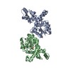
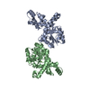
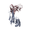
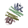
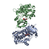
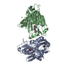
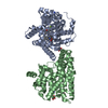
| Title | THE CRYSTAL STRUCTURE OF BACILLUS CEREUS PHOSPHONOACETALDEHYDE HYDROLASE COMPLEXED WITH TUNGSTATE, A PRODUCT ANALOG | ||||||
 Components Components | PHOSPHONOACETALDEHYDE HYDROLASE | ||||||
 Keywords Keywords | HYDROLASE / HAD-family alpha/beta core domain / Mg(II) binding site / 5-helix bundle | ||||||
| Function / homology |  Function and homology information Function and homology informationphosphonoacetaldehyde hydrolase / phosphonoacetaldehyde hydrolase activity / organic phosphonate catabolic process / phosphoglycolate phosphatase activity / DNA repair / magnesium ion binding / cytosol Similarity search - Function | ||||||
| Biological species |  | ||||||
| Method |  X-RAY DIFFRACTION / SYNCHROTRON / Resolution: 3 Å X-RAY DIFFRACTION / SYNCHROTRON / Resolution: 3 Å | ||||||
 Authors Authors | Morais, M.C. / Zhang, W. / Baker, A.S. / Zhang, G. / Dunaway-Mariano, D. / Allen, K.N. | ||||||
 Citation Citation | Journal: Biochemistry / Year: 2000 Title: The crystal structure of bacillus cereus phosphonoacetaldehyde hydrolase: insight into catalysis of phosphorus bond cleavage and catalytic diversification within the HAD enzyme superfamily. Authors: Morais, M.C. / Zhang, W. / Baker, A.S. / Zhang, G. / Dunaway-Mariano, D. / Allen, K.N. #1: Journal: Acta Crystallogr.,Sect.D / Year: 2000Title: Crystallization and Preliminary Crystallographic Analysis of Phosphonoacetaldehyde Hydrolase Authors: Morais, M.C. / Baker, A.S. / Dunaway-Mariano, D. / Allen, K.N. | ||||||
| History |
|
- Structure visualization
| Structure viewer | Molecule: MolmilJmol/JSmol |
|---|
- Downloads & links
-Download
| PDBx/mmCIF format | 1fez.cif.gz | 190.2 KB | Display | PDBx/mmCIF format |
|---|---|---|---|---|
| PDB format | pdb1fez.ent.gz | 155.1 KB | Display | PDB format |
| PDBx/mmJSON format | 1fez.json.gz | Tree view | PDBx/mmJSON format | |
| Others |  Other downloads Other downloads |
-Validation report
| Arichive directory | https://data.pdbj.org/pub/pdb/validation_reports/fe/1fezftp://data.pdbj.org/pub/pdb/validation_reports/fe/1fez | HTTPS FTP |
|---|
-Related structure data
| Similar structure data |
|---|


-Links
 PDBj
PDBj

- Assembly
| Deposited unit | 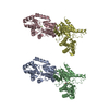
| ||||||||
|---|---|---|---|---|---|---|---|---|---|
| 1 | 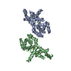
| ||||||||
| 2 | 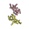
| ||||||||
| Unit cell |
| ||||||||
| Details | The biological assembly is a dimer constructed from chain A and chain B. The conformations of each monomer in the dimer are different. The crystallographic tetramer is generated by a non-crystallographic translation of this dimer |
-Components
| #1: Protein | Mass: 29162.453 Da / Num. of mol.: 4 Source method: isolated from a genetically manipulated source Source: (gene. exp.) #2: Chemical | ChemComp-MG /   Mass: 24.305 Da / Num. of mol.: 4 / Source method: obtained synthetically / Formula: Mg Mass: 24.305 Da / Num. of mol.: 4 / Source method: obtained synthetically / Formula: Mg#3: Chemical |   Mass: 247.838 Da / Num. of mol.: 2 / Source method: obtained synthetically / Formula: WO4 Mass: 247.838 Da / Num. of mol.: 2 / Source method: obtained synthetically / Formula: WO4#4: Water | ChemComp-HOH / |  Mass: 18.015 Da / Num. of mol.: 16 / Source method: isolated from a natural source / Formula: H2O Mass: 18.015 Da / Num. of mol.: 16 / Source method: isolated from a natural source / Formula: H2O |
|---|
-Experimental details
-Experiment
| Experiment | Method: X-RAY DIFFRACTION / Number of used crystals: 1 |
|---|
- Sample preparation
| Crystal | Density Matthews: 2.6 Å3/Da / Density % sol: 52.65 % | ||||||||||||||||||||||||||||||
|---|---|---|---|---|---|---|---|---|---|---|---|---|---|---|---|---|---|---|---|---|---|---|---|---|---|---|---|---|---|---|---|
| Crystal grow | Temperature: 291.14 K / Method: vapor diffusion, hanging drop / pH: 7.4 Details: PEG 4000, TRIS-HCl, magnesium chloride, , pH 7.4, VAPOR DIFFUSION, HANGING DROP, temperature 291.14K | ||||||||||||||||||||||||||||||
| Crystal grow | *PLUS | ||||||||||||||||||||||||||||||
| Components of the solutions | *PLUS
|
-Data collection
| Diffraction | Mean temperature: 93 K |
|---|---|
| Diffraction source | Source: SYNCHROTRON / Site: NSLS  / Beamline: X4A / Wavelength: 1.196994 / Beamline: X4A / Wavelength: 1.196994 |
| Detector | Type: FUJI / Detector: IMAGE PLATE / Date: Apr 30, 1997 |
| Radiation | Protocol: SINGLE WAVELENGTH / Monochromatic (M) / Laue (L): M / Scattering type: x-ray |
| Radiation wavelength | Wavelength: 1.196994 Å / Relative weight: 1 |
| Reflection | Resolution: 2.9→100 Å / Num. all: 38774 / Num. obs: 38774 / % possible obs: 93.7 % / Observed criterion σ(I): -1 / Redundancy: 2.42 % / Biso Wilson estimate: 16.54 Å2 / Rmerge(I) obs: 0.215 / Net I/σ(I): 6.56 |
| Reflection shell | Resolution: 3→100 Å / Rmerge(I) obs: 0.63 / Num. unique all: 3461 / % possible all: 91 |
| Reflection shell | *PLUS % possible obs: 91 % |
- Processing
| Software |
| |||||||||||||||||||||||||
|---|---|---|---|---|---|---|---|---|---|---|---|---|---|---|---|---|---|---|---|---|---|---|---|---|---|---|
| Refinement | Resolution: 3→100 Å / σ(F): 0 / Stereochemistry target values: Engh & HUBER
| |||||||||||||||||||||||||
| Refinement step | Cycle: LAST / Resolution: 3→100 Å
| |||||||||||||||||||||||||
| Refine LS restraints |
| |||||||||||||||||||||||||
| Software | *PLUS Name: CNS / Classification: refinement | |||||||||||||||||||||||||
| Refine LS restraints | *PLUS
|
