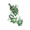
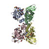
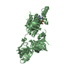
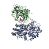
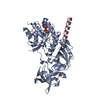
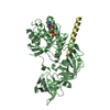
Entry Database : PDB / ID : 3wy9Title Crystal structure of a complex of the archaeal ribosomal stalk protein aP1 and the GDP-bound archaeal elongation factor aEF1alpha 50S ribosomal protein L12 Elongation factor 1-alpha Keywords / / / / / / / Function / homology Function Domain/homology Component
/ / / / / / / / / / / / / / / / / / / / / / / / / / / / / / / / / / / / / / / / / / / / Biological species Pyrococcus horikoshii OT3 (archaea)Method / / / Resolution : 2.3 Å Authors Ito, K. / Honda, T. / Suzuki, T. / Miyoshi, T. / Murakami, R. / Yao, M. / Uchiumi, T. Journal : Nucleic Acids Res. / Year : 2014Title : Molecular insights into the interaction of the ribosomal stalk protein with elongation factor 1 alpha.Authors : Ito, K. / Honda, T. / Suzuki, T. / Miyoshi, T. / Murakami, R. / Yao, M. / Uchiumi, T. History Deposition Aug 22, 2014 Deposition site / Processing site Revision 1.0 Dec 24, 2014 Provider / Type Revision 1.1 Aug 24, 2022 Group / Derived calculationsCategory citation / database_2 ... citation / database_2 / struct_ref_seq_dif / struct_site Item _citation.journal_volume / _citation.page_first ... _citation.journal_volume / _citation.page_first / _citation.page_last / _citation.title / _database_2.pdbx_DOI / _database_2.pdbx_database_accession / _struct_ref_seq_dif.details / _struct_site.pdbx_auth_asym_id / _struct_site.pdbx_auth_comp_id / _struct_site.pdbx_auth_seq_id Revision 1.2 May 29, 2024 Group / Category / chem_comp_bond
Show all Show less
 Movie
Movie Controller

 Yorodumi
 Open data
 Basic information
 Components
 Keywords
 Function and homology information
 Pyrococcus horikoshii OT3 (archaea)
 X-RAY DIFFRACTION /
  Authors
 Citation
 Structure visualization
 Downloads & links
 Other downloads
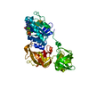


 PDBj
PDBj


 Assembly


 Type: RNA linking / Mass: 443.201 Da / Num. of mol.: 2 / Source method: obtained synthetically / Formula: C10H15N5O11P2 / Comment: GDP, energy-carrying molecule*YM
 Mass: 18.015 Da / Num. of mol.: 136 / Source method: isolated from a natural source / Formula: H2O
 Sample preparation
 / Beamline: BL-5A / Wavelength: 1 Å
 Processing