[English] 日本語
 Yorodumi
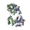
- PDB-1skq: The crystal structure of Sulfolobus solfataricus elongation facto... -
+ Open data
- Basic information
| Entry | Database: PDB / ID: 1skq | ||||||
|---|---|---|---|---|---|---|---|
| Title | The crystal structure of Sulfolobus solfataricus elongation factor 1-alpha in complex with magnesium and GDP | ||||||
 Components Components | Elongation factor 1-alpha | ||||||
 Keywords Keywords | TRANSLATION / Elongation factors / archaea / protein synthesis | ||||||
| Function / homology |  Function and homology information Function and homology informationprotein-synthesizing GTPase / translation elongation factor activity / GTPase activity / GTP binding / cytoplasm Similarity search - Function | ||||||
| Biological species |   Sulfolobus solfataricus (archaea) Sulfolobus solfataricus (archaea) | ||||||
| Method |  X-RAY DIFFRACTION / X-RAY DIFFRACTION /  SYNCHROTRON / SYNCHROTRON /  MOLECULAR REPLACEMENT / Resolution: 1.8 Å MOLECULAR REPLACEMENT / Resolution: 1.8 Å | ||||||
 Authors Authors | Vitagliano, L. / Ruggiero, A. / Masullo, M. / Cantiello, P. / Arcari, P. / Zagari, A. | ||||||
 Citation Citation |  Journal: Biochemistry / Year: 2004 Journal: Biochemistry / Year: 2004Title: The crystal structure of Sulfolobus solfataricus elongation factor 1alpha in complex with magnesium and GDP. Authors: Vitagliano, L. / Ruggiero, A. / Masullo, M. / Cantiello, P. / Arcari, P. / Zagari, A. #1:  Journal: Embo J. / Year: 2001 Journal: Embo J. / Year: 2001Title: THE CRYSTAL STRUCTURE OF SULFOLOBUS SOLFATARICUS ELONGATION FACTOR 1 ALPHA IN COMPLEX WITH GDP REVEALS NOVEL FEATURES IN NUCLEOTIDE BINDING AND EXCHANGE Authors: Vitagliano, L. / Masullo, M. / Sica, F. / Zagari, A. / Bocchini, V. | ||||||
| History |
| ||||||
| Remark 999 | SEQUENCE The sequence conflicts are due to strain differences between strains MT3 and MT4. The ...SEQUENCE The sequence conflicts are due to strain differences between strains MT3 and MT4. The protein crystallized is from strain MT4. The conflicts are noted in Swiss-Prot. |
- Structure visualization
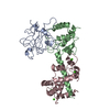
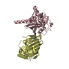
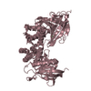
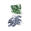
| Structure viewer | Molecule:  Molmil Molmil Jmol/JSmol Jmol/JSmol |
|---|
- Downloads & links
- Download
| PDBx/mmCIF format |  1skq.cif.gz 1skq.cif.gz | 182.5 KB | Display |  PDBx/mmCIF format PDBx/mmCIF format |
|---|---|---|---|---|
| PDB format |  pdb1skq.ent.gz pdb1skq.ent.gz | 143 KB | Display |  PDB format PDB format |
| PDBx/mmJSON format |  1skq.json.gz 1skq.json.gz | Tree view |  PDBx/mmJSON format PDBx/mmJSON format | |
| Others |  Other downloads Other downloads |
-Validation report
| Arichive directory |  https://data.pdbj.org/pub/pdb/validation_reports/sk/1skq https://data.pdbj.org/pub/pdb/validation_reports/sk/1skq ftp://data.pdbj.org/pub/pdb/validation_reports/sk/1skq ftp://data.pdbj.org/pub/pdb/validation_reports/sk/1skq | HTTPS FTP |
|---|
-Related structure data
| Related structure data | 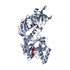 1jnyS S: Starting model for refinement |
|---|---|
| Similar structure data |
- Links
Links
- Assembly
| Deposited unit | 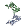
| ||||||||
|---|---|---|---|---|---|---|---|---|---|
| 1 | 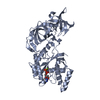
| ||||||||
| 2 | 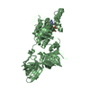
| ||||||||
| Unit cell |
| ||||||||
| Details | The biological assembly is the monomer |
- Components
| #1: Protein | Mass: 48539.059 Da / Num. of mol.: 2 Source method: isolated from a genetically manipulated source Source: (gene. exp.)   Sulfolobus solfataricus (archaea) / Strain: MT4 / Gene: TUF, TEF1, SSO0216 / Plasmid: pT7-7 / Species (production host): Escherichia coli / Production host: Sulfolobus solfataricus (archaea) / Strain: MT4 / Gene: TUF, TEF1, SSO0216 / Plasmid: pT7-7 / Species (production host): Escherichia coli / Production host:  #2: Chemical | ChemComp-MG / | #3: Chemical | #4: Water | ChemComp-HOH / | |
|---|
-Experimental details
-Experiment
| Experiment | Method:  X-RAY DIFFRACTION / Number of used crystals: 1 X-RAY DIFFRACTION / Number of used crystals: 1 |
|---|
- Sample preparation
| Crystal | Density Matthews: 2.94 Å3/Da / Density % sol: 58.21 % |
|---|---|
| Crystal grow | Temperature: 277 K / Method: microbatch under oil / pH: 5.6 Details: PEG4000, isopropanol, sodium citrate, pH 5.6, microbatch under oil, temperature 277K |
-Data collection
| Diffraction | Mean temperature: 100 K |
|---|---|
| Diffraction source | Source:  SYNCHROTRON / Site: SYNCHROTRON / Site:  ESRF ESRF  / Beamline: ID14-1 / Wavelength: 0.99 Å / Beamline: ID14-1 / Wavelength: 0.99 Å |
| Detector | Type: ADSC QUANTUM 4 / Detector: CCD / Date: Nov 30, 2001 |
| Radiation | Protocol: SINGLE WAVELENGTH / Monochromatic (M) / Laue (L): M / Scattering type: x-ray |
| Radiation wavelength | Wavelength: 0.99 Å / Relative weight: 1 |
| Reflection | Resolution: 1.8→30 Å / Num. all: 98914 / Num. obs: 98914 / % possible obs: 94.3 % / Observed criterion σ(F): 0 / Observed criterion σ(I): 0 / Rmerge(I) obs: 0.035 |
| Reflection shell | Resolution: 1.8→1.86 Å / Rmerge(I) obs: 0.343 / % possible all: 80 |
- Processing
| Software |
| ||||||||||||||||||||
|---|---|---|---|---|---|---|---|---|---|---|---|---|---|---|---|---|---|---|---|---|---|
| Refinement | Method to determine structure:  MOLECULAR REPLACEMENT MOLECULAR REPLACEMENTStarting model: PDB ENTRY 1JNY Resolution: 1.8→30 Å / Cross valid method: THROUGHOUT / σ(F): 0 / Stereochemistry target values: Engh & Huber
| ||||||||||||||||||||
| Refinement step | Cycle: LAST / Resolution: 1.8→30 Å
| ||||||||||||||||||||
| Refine LS restraints |
|
 Movie
Movie Controller




 PDBj
PDBj


