 Movie
Movie Controller

[English] 日本語
 Yorodumi
- PDB-4jpz: Voltage-gated sodium channel 1.2 C-terminal domain in complex wit... -
+ Open data

- Basic information
| Entry | Database: PDB / ID: 4jpz | ||||||
|---|---|---|---|---|---|---|---|
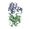
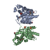
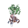
| Title | Voltage-gated sodium channel 1.2 C-terminal domain in complex with FGF13U and Ca2+/calmodulin | ||||||
 Components Components |
| ||||||
 Keywords Keywords | TRANSPORT PROTEIN / EF hand and IQ motif / ion channel / membrane | ||||||
| Function / homology |  Function and homology information Function and homology informationestablishment of neuroblast polarity / regulation of cardiac muscle cell action potential involved in regulation of contraction / negative regulation of collateral sprouting / positive regulation of voltage-gated sodium channel activity / intrinsic apoptotic signaling pathway in response to osmotic stress / : / : / : / : / positive regulation of protein autophosphorylation ...establishment of neuroblast polarity / regulation of cardiac muscle cell action potential involved in regulation of contraction / negative regulation of collateral sprouting / positive regulation of voltage-gated sodium channel activity / intrinsic apoptotic signaling pathway in response to osmotic stress / : / : / : / : / positive regulation of protein autophosphorylation / : / negative regulation of microtubule depolymerization / negative regulation of peptidyl-threonine phosphorylation / : / type 3 metabotropic glutamate receptor binding / cardiac muscle cell action potential involved in contraction / voltage-gated sodium channel complex / node of Ranvier / inhibitory synapse assembly / positive regulation of DNA binding / CaM pathway / Cam-PDE 1 activation / voltage-gated sodium channel activity / positive regulation of peptidyl-threonine phosphorylation / Sodium/Calcium exchangers / Interaction between L1 and Ankyrins / Calmodulin induced events / Reduction of cytosolic Ca++ levels / Activation of Ca-permeable Kainate Receptor / CREB1 phosphorylation through the activation of CaMKII/CaMKK/CaMKIV cascasde / Loss of phosphorylation of MECP2 at T308 / CREB1 phosphorylation through the activation of Adenylate Cyclase / negative regulation of high voltage-gated calcium channel activity / PKA activation / CaMK IV-mediated phosphorylation of CREB / Glycogen breakdown (glycogenolysis) / response to corticosterone / negative regulation of ryanodine-sensitive calcium-release channel activity / Activation of RAC1 downstream of NMDARs / organelle localization by membrane tethering / CLEC7A (Dectin-1) induces NFAT activation / : / autophagosome membrane docking / regulation of synaptic vesicle exocytosis / sodium ion transport / negative regulation of calcium ion export across plasma membrane / regulation of cardiac muscle cell action potential / presynaptic endocytosis / Synthesis of IP3 and IP4 in the cytosol / cerebral cortex cell migration / positive regulation of protein serine/threonine kinase activity / Phase 0 - rapid depolarisation / Negative regulation of NMDA receptor-mediated neuronal transmission / microtubule polymerization / Unblocking of NMDA receptors, glutamate binding and activation / calcineurin-mediated signaling / RHO GTPases activate PAKs / nitric-oxide synthase binding / regulation of cell communication by electrical coupling involved in cardiac conduction / Ion transport by P-type ATPases / adenylate cyclase binding / Uptake and function of anthrax toxins / lateral plasma membrane / protein phosphatase activator activity / intercalated disc / regulation of ryanodine-sensitive calcium-release channel activity / Long-term potentiation / sodium channel regulator activity / Calcineurin activates NFAT / Regulation of MECP2 expression and activity / DARPP-32 events / Smooth Muscle Contraction / regulation of synaptic vesicle endocytosis / detection of calcium ion / catalytic complex / regulation of cardiac muscle contraction / cellular response to interferon-beta / RHO GTPases activate IQGAPs / positive regulation of nitric-oxide synthase activity / phosphatidylinositol 3-kinase binding / beta-tubulin binding / activation of adenylate cyclase activity / calcium channel inhibitor activity / presynaptic cytosol / neuronal action potential / Activation of AMPK downstream of NMDARs / regulation of release of sequestered calcium ion into cytosol by sarcoplasmic reticulum / enzyme regulator activity / eNOS activation / Ion homeostasis / Tetrahydrobiopterin (BH4) synthesis, recycling, salvage and regulation / regulation of calcium-mediated signaling / Protein methylation / titin binding / regulation of cardiac muscle contraction by regulation of the release of sequestered calcium ion / myelination / voltage-gated potassium channel complex / neurogenesis / FCERI mediated Ca+2 mobilization / calcium channel complex Similarity search - Function | ||||||
| Biological species |  Homo sapiens (human) Homo sapiens (human) | ||||||
| Method |  X-RAY DIFFRACTION / SYNCHROTRON / SAD / Resolution: 3.02 Å X-RAY DIFFRACTION / SYNCHROTRON / SAD / Resolution: 3.02 Å | ||||||
 Authors Authors | Wang, C. / Chung, B.C. / Yan, H. / Wang, H.G. / Lee, S.Y. / Pitt, G.S. | ||||||
 Citation Citation | Journal: Nat Commun / Year: 2014 Title: Structural analyses of Ca(2+)/CaM interaction with NaV channel C-termini reveal mechanisms of calcium-dependent regulation. Authors: Wang, C. / Chung, B.C. / Yan, H. / Wang, H.G. / Lee, S.Y. / Pitt, G.S. | ||||||
| History |
|
- Structure visualization
| Structure viewer | Molecule: MolmilJmol/JSmol |
|---|
- Downloads & links
-Download
| PDBx/mmCIF format | 4jpz.cif.gz | 187.4 KB | Display | PDBx/mmCIF format |
|---|---|---|---|---|
| PDB format | pdb4jpz.ent.gz | 148 KB | Display | PDB format |
| PDBx/mmJSON format | 4jpz.json.gz | Tree view | PDBx/mmJSON format | |
| Others |  Other downloads Other downloads |
-Validation report
| Arichive directory | https://data.pdbj.org/pub/pdb/validation_reports/jp/4jpzftp://data.pdbj.org/pub/pdb/validation_reports/jp/4jpz | HTTPS FTP |
|---|
-Related structure data


-Links
 PDBj
PDBj


- Assembly
| Deposited unit | 
| ||||||||
|---|---|---|---|---|---|---|---|---|---|
| 1 | 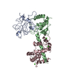
| ||||||||
| 2 | 
| ||||||||
| Unit cell |
|
-Components
| #1: Protein | Mass: 21609.592 Da / Num. of mol.: 2 Source method: isolated from a genetically manipulated source Source: (gene. exp.) Homo sapiens (human) / Gene: FGF13, FHF2 / Production host:  #2: Protein | Mass: 21036.049 Da / Num. of mol.: 2 Source method: isolated from a genetically manipulated source Source: (gene. exp.) Homo sapiens (human) / Gene: SCN2A, NAC2, SCN2A1, SCN2A2 / Production host: #3: Protein | Mass: 16852.545 Da / Num. of mol.: 2 Source method: isolated from a genetically manipulated source Source: (gene. exp.) Homo sapiens (human)Gene: CALM1, CALM, CAM, CAM1, CALM2, CAM2, CAMB, CALM3, CALML2, CAM3, CAMC, CAMIII Production host: #4: Chemical | ChemComp-CA /   Mass: 40.078 Da / Num. of mol.: 8 / Source method: obtained synthetically / Formula: Ca Mass: 40.078 Da / Num. of mol.: 8 / Source method: obtained synthetically / Formula: Ca#5: Water | ChemComp-HOH / |  Mass: 18.015 Da / Num. of mol.: 31 / Source method: isolated from a natural source / Formula: H2O Mass: 18.015 Da / Num. of mol.: 31 / Source method: isolated from a natural source / Formula: H2OSequence details | THE RESIDUES RRRP AT POSITIONS 59-62 ARE ISOFORM 2 NATURAL VARIANTS, UNP CODE Q92913-2 | |
|---|
-Experimental details
-Experiment
| Experiment | Method: X-RAY DIFFRACTION / Number of used crystals: 1 |
|---|
- Sample preparation
| Crystal | Density Matthews: 2.98 Å3/Da / Density % sol: 58.67 % |
|---|---|
| Crystal grow | Temperature: 290 K / Method: evaporation / pH: 7.5 Details: 14% pEG3350, 300 mM sodium acetate,50 mM Tris pH 7.5, and 2 mM CaCl2, EVAPORATION, temperature 290K |
-Data collection
| Diffraction | Mean temperature: 100 K | |||||||||||||||||||||||||||||||||||||||||||||
|---|---|---|---|---|---|---|---|---|---|---|---|---|---|---|---|---|---|---|---|---|---|---|---|---|---|---|---|---|---|---|---|---|---|---|---|---|---|---|---|---|---|---|---|---|---|---|
| Diffraction source | Source: SYNCHROTRON / Site: APS  / Beamline: 24-ID-C / Wavelength: 0.97 Å / Beamline: 24-ID-C / Wavelength: 0.97 Å | |||||||||||||||||||||||||||||||||||||||||||||
| Detector | Type: ADSC QUANTUM 315 / Detector: CCD / Date: Aug 12, 2012 | |||||||||||||||||||||||||||||||||||||||||||||
| Radiation | Monochromator: double crystal, Si 111 / Protocol: SINGLE WAVELENGTH / Monochromatic (M) / Laue (L): M / Scattering type: x-ray | |||||||||||||||||||||||||||||||||||||||||||||
| Radiation wavelength | Wavelength: 0.97 Å / Relative weight: 1 | |||||||||||||||||||||||||||||||||||||||||||||
| Reflection | Resolution: 3.02→50 Å / Num. all: 53782 / Num. obs: 53782 / % possible obs: 99.8 % / Observed criterion σ(F): 0 / Observed criterion σ(I): -3 | |||||||||||||||||||||||||||||||||||||||||||||
| Reflection shell |
|
- Processing
| Software |
| |||||||||||||||||||||||||||||||||||||||||||||||||||||||||||||||||||||||||||||
|---|---|---|---|---|---|---|---|---|---|---|---|---|---|---|---|---|---|---|---|---|---|---|---|---|---|---|---|---|---|---|---|---|---|---|---|---|---|---|---|---|---|---|---|---|---|---|---|---|---|---|---|---|---|---|---|---|---|---|---|---|---|---|---|---|---|---|---|---|---|---|---|---|---|---|---|---|---|---|
| Refinement | Method to determine structure: SAD / Resolution: 3.02→48.23 Å / SU ML: 0.44 / σ(F): 1.4 / Phase error: 28.43 / Stereochemistry target values: ML
| |||||||||||||||||||||||||||||||||||||||||||||||||||||||||||||||||||||||||||||
| Solvent computation | Shrinkage radii: 0.9 Å / VDW probe radii: 1.11 Å / Solvent model: FLAT BULK SOLVENT MODEL | |||||||||||||||||||||||||||||||||||||||||||||||||||||||||||||||||||||||||||||
| Refinement step | Cycle: LAST / Resolution: 3.02→48.23 Å
| |||||||||||||||||||||||||||||||||||||||||||||||||||||||||||||||||||||||||||||
| Refine LS restraints |
| |||||||||||||||||||||||||||||||||||||||||||||||||||||||||||||||||||||||||||||
| LS refinement shell |
|
