- PDB-3sc3: Crystal structure of a Putative DNA replication regulator Hda (Sa... -
+
Open data
ID or keywords:
Loading...
-
Basic information
Entry
Database: PDB / ID: 3sc3
Title
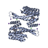
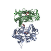
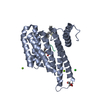
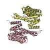
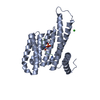
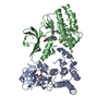
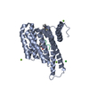
Crystal structure of a Putative DNA replication regulator Hda (Sama_1916) from SHEWANELLA AMAZONENSIS SB2B at 3.00 A resolution
Components
Putative DNA replication regulator Hda
Keywords
HYDROLASE REGULATOR / DNA BINDING PROTEIN / P-loop containing nucleoside triphosphate hydrolases / Structural Genomics / Joint Center for Structural Genomics / JCSG / Protein Structure Initiative / PSI-BIOLOGY / HYDROLASE
Function / homology
Function and homology information
DNA replication initiation / negative regulation of DNA-templated DNA replication initiation / nucleotide binding / metal ion binding Similarity search - Function
ANALYTICAL SIZE EXCLUSION CHROMATOGRAPHY WITH STATIC LIGHT SCATTERING SUPPORTS THE ASSIGNMENT OF A DIMER AS A SIGNIFICANT OLIGOMERIZATION STATE IN SOLUTION.
-
Components
#1: Protein
PutativeDNAreplicationregulatorHda / Regulatory inactivation of DnaA Hda protein
Mass: 25509.047 Da / Num. of mol.: 2 Source method: isolated from a genetically manipulated source Source: (gene. exp.) Shewanella amazonensis (bacteria) / Strain: SB2B / Gene: SAMA_14OCT04_CONTIG53_REVISED_GENE1514, Sama_1916 / Plasmid: SpeedET / Production host: Escherichia Coli (E. coli) / Strain (production host): HK100 / References: UniProt: A1S6W5
Mass: 96.063 Da / Num. of mol.: 5 / Source method: obtained synthetically / Formula: SO4
Has protein modification
Y
Sequence details
THE CONSTRUCT (RESIDUES 18-241) WAS EXPRESSED WITH A PURIFICATION TAG MGSDKIHHHHHHENLYFQG. THE TAG ...THE CONSTRUCT (RESIDUES 18-241) WAS EXPRESSED WITH A PURIFICATION TAG MGSDKIHHHHHHENLYFQG. THE TAG WAS REMOVED WITH TEV PROTEASE LEAVING ONLY A GLYCINE (0) FOLLOWED BY THE TARGET SEQUENCE.
-
Experimental details
-
Experiment
Experiment
Method: X-RAY DIFFRACTION / Number of used crystals: 1
-
Sample preparation
Crystal
Density Matthews: 3.5 Å3/Da / Density % sol: 64.86 %
Crystal grow
Temperature: 277 K / Method: vapor diffusion, sitting drop / pH: 7.5 Details: 1.26M (NH4)2SO4, 0.1M HEPES pH 7.5, NANODROP, VAPOR DIFFUSION, SITTING DROP, temperature 277K
Type: MARMOSAIC 325 mm CCD / Detector: CCD / Date: Nov 15, 2008 Details: Flat mirror (vertical focusing); single crystal Si(111) bent monochromator (ho rizontal focusing)
Radiation
Monochromator: single crystal Si(111) bent / Protocol: SINGLE WAVELENGTH / Monochromatic (M) / Laue (L): M / Scattering type: x-ray
Radiation wavelength
Wavelength: 0.97799 Å / Relative weight: 1
Reflection
Resolution: 3→46.881 Å / Num. obs: 14422 / % possible obs: 99.9 % / Observed criterion σ(I): -3 / Biso Wilson estimate: 91.955 Å2 / Rmerge(I) obs: 0.143 / Net I/σ(I): 10.68
Reflection shell
Rmerge(I) obs: 0.014 / Diffraction-ID: 1
Resolution (Å)
Highest resolution (Å)
Mean I/σ(I) obs
Num. measured obs
Num. unique obs
% possible all
3-3.17
1.6
16285
2175
99.9
3.17-3.28
2.7
8745
1163
100
3.28-3.41
3.6
9113
1211
100
3.41-3.57
4.9
9140
1222
100
3.57-3.76
7.5
9312
1239
100
3.76-3.99
9.8
8967
1199
100
3.99-4.29
13.1
8984
1200
99.9
4.29-4.72
16.3
9219
1230
100
4.72-5.39
18.2
9077
1218
100
5.39-6.75
17.7
9191
1246
100
6.75
27.2
9314
1324
99.4
-
Phasing
Phasing
Method: SAD
-
Processing
Software
Name
Version
Classification
NB
MolProbity
3beta29
modelbuilding
PDB_EXTRACT
3.1
dataextraction
SHELX
phasing
SHARP
phasing
XSCALE
December6, 2007
datascaling
PHENIX
1.4
refinement
XDS
datareduction
SHELXD
phasing
Refinement
Method to determine structure: SAD / Resolution: 3.001→46.881 Å / Occupancy max: 1 / Occupancy min: 0.66 / SU ML: 1.17 / σ(F): 1.39 / Phase error: 27.67 / Stereochemistry target values: MLHL Details: 1. A MET-INHIBITION PROTOCOL WAS USED FOR SELENOMETHIONINE INCORPORATION DURING PROTEIN EXPRESSION. THE OCCUPANCY OF THE SE ATOMS IN THE MSE RESIDUES WAS REDUCED TO 0.75 FOR THE REDUCED ...Details: 1. A MET-INHIBITION PROTOCOL WAS USED FOR SELENOMETHIONINE INCORPORATION DURING PROTEIN EXPRESSION. THE OCCUPANCY OF THE SE ATOMS IN THE MSE RESIDUES WAS REDUCED TO 0.75 FOR THE REDUCED SCATTERING POWER DUE TO PARTIAL S-MET INCORPORATION. 2. SULFATE (SO4) MODELED ARE PRESENT IN CRYSTLLIZATION/CRYO CONDITION.
Rfactor
Num. reflection
% reflection
Rfree
0.2532
1449
10.06 %
Rwork
0.2237
-
-
obs
0.2267
14406
99.9 %
Solvent computation
Shrinkage radii: 0.9 Å / VDW probe radii: 1.11 Å / Solvent model: FLAT BULK SOLVENT MODEL / Bsol: 83.041 Å2 / ksol: 0.351 e/Å3
In the structure databanks used in Yorodumi, some data are registered as the other names, "COVID-19 virus" and "2019-nCoV". Here are the details of the virus and the list of structure data.
Jan 31, 2019. EMDB accession codes are about to change! (news from PDBe EMDB page)
EMDB accession codes are about to change! (news from PDBe EMDB page)
The allocation of 4 digits for EMDB accession codes will soon come to an end. Whilst these codes will remain in use, new EMDB accession codes will include an additional digit and will expand incrementally as the available range of codes is exhausted. The current 4-digit format prefixed with “EMD-” (i.e. EMD-XXXX) will advance to a 5-digit format (i.e. EMD-XXXXX), and so on. It is currently estimated that the 4-digit codes will be depleted around Spring 2019, at which point the 5-digit format will come into force.
The EM Navigator/Yorodumi systems omit the EMD- prefix.
Related info.:Q: What is EMD? / ID/Accession-code notation in Yorodumi/EM Navigator
Yorodumi is a browser for structure data from EMDB, PDB, SASBDB, etc.
This page is also the successor to EM Navigator detail page, and also detail information page/front-end page for Omokage search.
The word "yorodu" (or yorozu) is an old Japanese word meaning "ten thousand". "mi" (miru) is to see.
Related info.:EMDB / PDB / SASBDB / Comparison of 3 databanks / Yorodumi Search / Aug 31, 2016. New EM Navigator & Yorodumi / Yorodumi Papers / Jmol/JSmol / Function and homology information / Changes in new EM Navigator and Yorodumi
 Movie
Movie Controller

 Yorodumi
 Open data
 Basic information
 Components
 Keywords
 Function and homology information
 Shewanella amazonensis (bacteria)
 X-RAY DIFFRACTION /
  Authors
 Citation
 Structure visualization
 Downloads & links
 Other downloads


 PDBj
PDBj
 Assembly
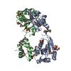

 Mass: 96.063 Da / Num. of mol.: 5 / Source method: obtained synthetically / Formula: SO4
 Sample preparation
 / Beamline: BL11-1 / Wavelength: 0.97799
  Processing