 ムービー
ムービー コントローラー


+ データを開く

- 基本情報
基本情報
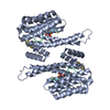
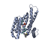
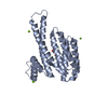
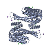
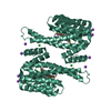
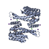
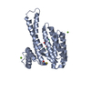
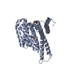
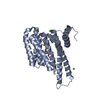
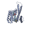
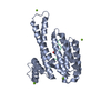
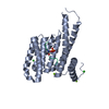
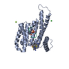
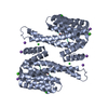
| 登録情報 | データベース: PDB / ID: 6rkk | ||||||
|---|---|---|---|---|---|---|---|
| タイトル | Fragment AZ-021 binding at the p53pT387/14-3-3 sigma interface | ||||||
 要素 要素 |
| ||||||
 キーワード キーワード | PEPTIDE BINDING PROTEIN / protein protein interaction / fragment soaking / stabilization | ||||||
| 機能・相同性 |  機能・相同性情報 機能・相同性情報negative regulation of helicase activity / Loss of function of TP53 in cancer due to loss of tetramerization ability / Regulation of TP53 Expression / signal transduction by p53 class mediator / negative regulation of G1 to G0 transition / negative regulation of glucose catabolic process to lactate via pyruvate / Transcriptional activation of cell cycle inhibitor p21 / regulation of intrinsic apoptotic signaling pathway by p53 class mediator / negative regulation of pentose-phosphate shunt / Activation of NOXA and translocation to mitochondria ...negative regulation of helicase activity / Loss of function of TP53 in cancer due to loss of tetramerization ability / Regulation of TP53 Expression / signal transduction by p53 class mediator / negative regulation of G1 to G0 transition / negative regulation of glucose catabolic process to lactate via pyruvate / Transcriptional activation of cell cycle inhibitor p21 / regulation of intrinsic apoptotic signaling pathway by p53 class mediator / negative regulation of pentose-phosphate shunt / Activation of NOXA and translocation to mitochondria / ATP-dependent DNA/DNA annealing activity / regulation of cell cycle G2/M phase transition / oligodendrocyte apoptotic process / negative regulation of miRNA processing / intrinsic apoptotic signaling pathway in response to hypoxia / oxidative stress-induced premature senescence / regulation of tissue remodeling / positive regulation of thymocyte apoptotic process / positive regulation of mitochondrial membrane permeability / germ cell nucleus / regulation of fibroblast apoptotic process / bone marrow development / cellular response to actinomycin D / circadian behavior / regulation of mitochondrial membrane permeability involved in apoptotic process / histone deacetylase regulator activity / positive regulation of programmed necrotic cell death / : / RUNX3 regulates CDKN1A transcription / T cell proliferation involved in immune response / TP53 Regulates Transcription of Death Receptors and Ligands / Activation of PUMA and translocation to mitochondria / TP53 regulates transcription of additional cell cycle genes whose exact role in the p53 pathway remain uncertain / mRNA transcription / negative regulation of glial cell proliferation / negative regulation of neuroblast proliferation / regulation of DNA damage response, signal transduction by p53 class mediator / Regulation of TP53 Activity through Association with Co-factors / Formation of Senescence-Associated Heterochromatin Foci (SAHF) / mitochondrial DNA repair / T cell lineage commitment / thymocyte apoptotic process / ER overload response / positive regulation of epidermal cell differentiation / keratinocyte development / TP53 Regulates Transcription of Caspase Activators and Caspases / cardiac septum morphogenesis / regulation of epidermal cell division / necroptotic process / protein kinase C inhibitor activity / B cell lineage commitment / keratinization / entrainment of circadian clock by photoperiod / negative regulation of DNA replication / Zygotic genome activation (ZGA) / negative regulation of mitophagy / TP53 Regulates Transcription of Genes Involved in Cytochrome C Release / PI5P Regulates TP53 Acetylation / positive regulation of release of cytochrome c from mitochondria / regulation of cell-cell adhesion / neuroblast proliferation / Association of TriC/CCT with target proteins during biosynthesis / negative regulation of telomere maintenance via telomerase / SUMOylation of transcription factors / TP53 regulates transcription of several additional cell death genes whose specific roles in p53-dependent apoptosis remain uncertain / keratinocyte proliferation / rRNA transcription / establishment of skin barrier / negative regulation of reactive oxygen species metabolic process / intrinsic apoptotic signaling pathway by p53 class mediator / TFIID-class transcription factor complex binding / Transcriptional Regulation by VENTX / Regulation of localization of FOXO transcription factors / cellular response to UV-C / replicative senescence / viral process / intrinsic apoptotic signaling pathway in response to endoplasmic reticulum stress / hematopoietic stem cell differentiation / negative regulation of keratinocyte proliferation / embryonic organ development / phosphoserine residue binding / Activation of BAD and translocation to mitochondria / intrinsic apoptotic signaling pathway in response to DNA damage by p53 class mediator / Pyroptosis / positive regulation of RNA polymerase II transcription preinitiation complex assembly / chromosome organization / general transcription initiation factor binding / positive regulation of execution phase of apoptosis / type II interferon-mediated signaling pathway / hematopoietic progenitor cell differentiation / cAMP/PKA signal transduction / negative regulation of stem cell proliferation / negative regulation of protein localization to plasma membrane / TP53 Regulates Transcription of Genes Involved in G1 Cell Cycle Arrest / response to X-ray / somitogenesis / SARS-CoV-2 targets host intracellular signalling and regulatory pathways / negative regulation of fibroblast proliferation / negative regulation of protein kinase activity / core promoter sequence-specific DNA binding 類似検索 - 分子機能 | ||||||
| 生物種 |  Homo sapiens (ヒト) Homo sapiens (ヒト) | ||||||
| 手法 |  X線回折 / 分子置換 / 解像度: 1.88 Å X線回折 / 分子置換 / 解像度: 1.88 Å | ||||||
 データ登録者 データ登録者 | Genet, S. / Wolter, M. / Guillory, X. / Somsen, B. / Leysen, S. / Patel, J. / Castaldi, P. / Ottmann, C. | ||||||
| 資金援助 |  オランダ, 1件 オランダ, 1件
| ||||||
 引用 引用 | ジャーナル: J.Med.Chem. / 年: 2020 タイトル: Fragment-based Differential Targeting of PPI Stabilizer Interfaces. 著者: Guillory, X. / Wolter, M. / Leysen, S. / Neves, J.F. / Kuusk, A. / Genet, S. / Somsen, B. / Morrow, J.K. / Rivers, E. / van Beek, L. / Patel, J. / Goodnow, R. / Schoenherr, H. / Fuller, N. / ...著者: Guillory, X. / Wolter, M. / Leysen, S. / Neves, J.F. / Kuusk, A. / Genet, S. / Somsen, B. / Morrow, J.K. / Rivers, E. / van Beek, L. / Patel, J. / Goodnow, R. / Schoenherr, H. / Fuller, N. / Cao, Q. / Doveston, R.G. / Brunsveld, L. / Arkin, M.R. / Castaldi, P. / Boyd, H. / Landrieu, I. / Chen, H. / Ottmann, C. | ||||||
| 履歴 |
|
- 構造の表示
構造の表示
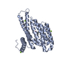
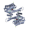
| 構造ビューア | 分子: MolmilJmol/JSmol |
|---|
- ダウンロードとリンク
-ダウンロード
| PDBx/mmCIF形式 | 6rkk.cif.gz | 114.2 KB | 表示 | PDBx/mmCIF形式 |
|---|---|---|---|---|
| PDB形式 | pdb6rkk.ent.gz | 86.7 KB | 表示 | PDB形式 |
| PDBx/mmJSON形式 | 6rkk.json.gz | ツリー表示 | PDBx/mmJSON形式 | |
| その他 |  その他のダウンロード その他のダウンロード |
-検証レポート
| アーカイブディレクトリ | https://data.pdbj.org/pub/pdb/validation_reports/rk/6rkkftp://data.pdbj.org/pub/pdb/validation_reports/rk/6rkk | HTTPS FTP |
|---|
-関連構造データ
| 関連構造データ | 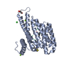 6r5lC  6rhcC  6rjlC  6rjqC 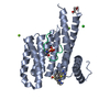 6rjzC  6rk8C  6rkiC 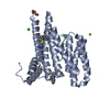 6rkmC 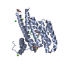 6rl3C 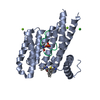 6rl4C  6rl6C  6rm5C 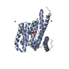 6rm7C  6rp6C 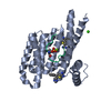 6rwhC  6rwiC 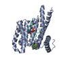 6rwsC 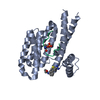 6rwuC 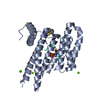 6rx2C 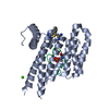 6s39C 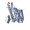 6s3cC  6s40C 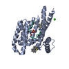 6s9qC 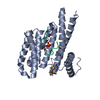 6sinC 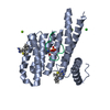 6sioC 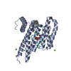 6sipC 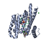 6siqC 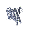 6slvC  6slwC  6slxC 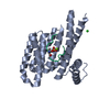 5mocS S: 精密化の開始モデル C: 同じ文献を引用 ( |
|---|---|
| 類似構造データ |


-リンク
 PDBj
PDBj


- 集合体
集合体
| 登録構造単位 | 
| ||||||||
|---|---|---|---|---|---|---|---|---|---|
| 1 | 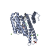
| ||||||||
| 単位格子 |
|
-要素
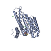
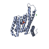
-タンパク質 / タンパク質・ペプチド , 2種, 2分子 AP
| #1: タンパク質 | 分子量: 28210.518 Da / 分子数: 1 / 由来タイプ: 組換発現 / 詳細: Residues -4 to 0 are expression tag / 由来: (組換発現) Homo sapiens (ヒト) / 遺伝子: SFN, HME1 / 発現宿主:  |
|---|---|
| #2: タンパク質・ペプチド | 分子量: 1449.520 Da / 分子数: 1 / 由来タイプ: 合成 / 由来: (合成) Homo sapiens (ヒト) / 参照: UniProt: P04637 |
-非ポリマー , 5種, 392分子 


| #3: 化合物 |  分子量: 24.305 Da / 分子数: 2 / 由来タイプ: 合成 / 式: Mg 分子量: 24.305 Da / 分子数: 2 / 由来タイプ: 合成 / 式: Mg#4: 化合物 | ChemComp-CL / |  分子量: 35.453 Da / 分子数: 1 / 由来タイプ: 合成 / 式: Cl 分子量: 35.453 Da / 分子数: 1 / 由来タイプ: 合成 / 式: Cl#5: 化合物 | ChemComp-GOL / |  分子量: 92.094 Da / 分子数: 1 / 由来タイプ: 合成 / 式: C3H8O3 分子量: 92.094 Da / 分子数: 1 / 由来タイプ: 合成 / 式: C3H8O3#6: 化合物 | ChemComp-K6W / |  分子量: 292.398 Da / 分子数: 1 / 由来タイプ: 合成 / 式: C18H16N2S 分子量: 292.398 Da / 分子数: 1 / 由来タイプ: 合成 / 式: C18H16N2S#7: 水 | ChemComp-HOH / | 分子量: 18.015 Da / 分子数: 387 / 由来タイプ: 天然 / 式: H2O |
|---|
-詳細
| Has protein modification | Y |
|---|
-実験情報
-実験
| 実験 | 手法: X線回折 / 使用した結晶の数: 1 |
|---|
- 試料調製
試料調製
| 結晶 | マシュー密度: 2.44 Å3/Da / 溶媒含有率: 49.52 % |
|---|---|
| 結晶化 | 温度: 277 K / 手法: 蒸気拡散法, ハンギングドロップ法 / pH: 7.5 詳細: 0.1M Hepes, pH7.5, 27%PEG, 5% Glycerol, 0.2M Calcium Chloride, 2mM DTT. |
-データ収集
| 回折 | 平均測定温度: 100 K / Serial crystal experiment: N |
|---|---|
| 放射光源 | 由来: SEALED TUBE / タイプ: RIGAKU MICROMAX-003 / 波長: 1.54187 Å |
| 検出器 | タイプ: DECTRIS PILATUS 200K / 検出器: PIXEL / 日付: 2017年8月9日 |
| 放射 | プロトコル: SINGLE WAVELENGTH / 単色(M)・ラウエ(L): M / 散乱光タイプ: x-ray |
| 放射波長 | 波長: 1.54187 Å / 相対比: 1 |
| 反射 | 解像度: 1.88→26.6 Å / Num. obs: 23784 / % possible obs: 99.9 % / 冗長度: 4.9 % / CC1/2: 0.947 / Rrim(I) all: 0.276 / Net I/σ(I): 4.2 |
| 反射 シェル | 解像度: 1.88→1.93 Å / 冗長度: 4.6 % / Mean I/σ(I) obs: 1.7 / Num. unique obs: 1479 / CC1/2: 0.562 / Rrim(I) all: 0.861 / % possible all: 100 |
- 解析
解析
| ソフトウェア |
| |||||||||||||||||||||||||||||||||||||||||||||||||||||||||||||||
|---|---|---|---|---|---|---|---|---|---|---|---|---|---|---|---|---|---|---|---|---|---|---|---|---|---|---|---|---|---|---|---|---|---|---|---|---|---|---|---|---|---|---|---|---|---|---|---|---|---|---|---|---|---|---|---|---|---|---|---|---|---|---|---|---|
| 精密化 | 構造決定の手法: 分子置換 開始モデル: 5MOC 解像度: 1.88→25.64 Å / SU ML: 0.24 / 交差検証法: FREE R-VALUE / σ(F): 1.33 / 位相誤差: 26.8
| |||||||||||||||||||||||||||||||||||||||||||||||||||||||||||||||
| 溶媒の処理 | 減衰半径: 0.9 Å / VDWプローブ半径: 1.11 Å | |||||||||||||||||||||||||||||||||||||||||||||||||||||||||||||||
| 精密化ステップ | サイクル: LAST / 解像度: 1.88→25.64 Å
| |||||||||||||||||||||||||||||||||||||||||||||||||||||||||||||||
| 拘束条件 |
| |||||||||||||||||||||||||||||||||||||||||||||||||||||||||||||||
| LS精密化 シェル |
|
