 Movie
Movie Controller

[English] 日本語
 Yorodumi
- PDB-6zcj: 14-3-3sigma in complex with SLP76pS376 phosphopeptide crystal str... -
+ Open data

- Basic information
| Entry | Database: PDB / ID: 6zcj | ||||||
|---|---|---|---|---|---|---|---|
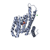
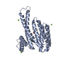
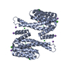
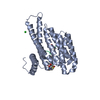
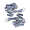
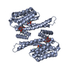
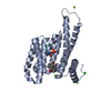
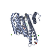
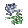
| Title | 14-3-3sigma in complex with SLP76pS376 phosphopeptide crystal structure | ||||||
 Components Components |
| ||||||
 Keywords Keywords | PEPTIDE BINDING PROTEIN / Adaptor Protein / Phosphorylation | ||||||
| Function / homology |  Function and homology information Function and homology informationTCR signalosome / mast cell activation / positive regulation of epidermal cell differentiation / keratinocyte development / regulation of epidermal cell division / protein kinase C inhibitor activity / positive regulation of protein kinase activity / keratinization / regulation of cell-cell adhesion / keratinocyte proliferation ...TCR signalosome / mast cell activation / positive regulation of epidermal cell differentiation / keratinocyte development / regulation of epidermal cell division / protein kinase C inhibitor activity / positive regulation of protein kinase activity / keratinization / regulation of cell-cell adhesion / keratinocyte proliferation / establishment of skin barrier / Generation of second messenger molecules / Regulation of localization of FOXO transcription factors / negative regulation of keratinocyte proliferation / phosphoserine residue binding / Activation of BAD and translocation to mitochondria / plasma membrane raft / cAMP/PKA signal transduction / negative regulation of stem cell proliferation / negative regulation of protein localization to plasma membrane / SARS-CoV-2 targets host intracellular signalling and regulatory pathways / negative regulation of protein kinase activity / Chk1/Chk2(Cds1) mediated inactivation of Cyclin B:Cdk1 complex / SARS-CoV-1 targets host intracellular signalling and regulatory pathways / RHO GTPases activate PKNs / GPVI-mediated activation cascade / positive regulation of protein localization / stem cell proliferation / protein export from nucleus / FCERI mediated Ca+2 mobilization / release of cytochrome c from mitochondria / TP53 Regulates Transcription of Genes Involved in G2 Cell Cycle Arrest / negative regulation of innate immune response / positive regulation of cell adhesion / cell surface receptor protein tyrosine kinase signaling pathway / positive regulation of protein export from nucleus / TP53 Regulates Metabolic Genes / Translocation of SLC2A4 (GLUT4) to the plasma membrane / FCERI mediated MAPK activation / protein sequestering activity / intrinsic apoptotic signaling pathway in response to DNA damage / cell-cell junction / intracellular protein localization / DAP12 signaling / T cell receptor signaling pathway / regulation of protein localization / sperm midpiece / positive regulation of cell growth / regulation of cell cycle / intracellular signal transduction / immune response / cadherin binding / protein kinase binding / negative regulation of transcription by RNA polymerase II / signal transduction / : / extracellular exosome / identical protein binding / nucleus / cytosol / cytoplasm Similarity search - Function | ||||||
| Biological species |  Homo sapiens (human) Homo sapiens (human)synthetic construct (others) | ||||||
| Method |  X-RAY DIFFRACTION / SYNCHROTRON / MOLECULAR REPLACEMENT / Resolution: 1.53 Å X-RAY DIFFRACTION / SYNCHROTRON / MOLECULAR REPLACEMENT / Resolution: 1.53 Å | ||||||
 Authors Authors | Soini, L. / Leysen, S. / Davis, J. / Ottmann, C. | ||||||
| Funding support |  Netherlands, 1items Netherlands, 1items
| ||||||
 Citation Citation | Journal: Febs Lett. / Year: 2021 Title: The 14-3-3/SLP76 protein-protein interaction in T-cell receptor signalling: a structural and biophysical characterization. Authors: Soini, L. / Leysen, S. / Davis, J. / Westwood, M. / Ottmann, C. | ||||||
| History |
|
- Structure visualization
| Structure viewer | Molecule: MolmilJmol/JSmol |
|---|
- Downloads & links
-Download
| PDBx/mmCIF format | 6zcj.cif.gz | 88.8 KB | Display | PDBx/mmCIF format |
|---|---|---|---|---|
| PDB format | pdb6zcj.ent.gz | 53.1 KB | Display | PDB format |
| PDBx/mmJSON format | 6zcj.json.gz | Tree view | PDBx/mmJSON format | |
| Others |  Other downloads Other downloads |
-Validation report
| Arichive directory | https://data.pdbj.org/pub/pdb/validation_reports/zc/6zcjftp://data.pdbj.org/pub/pdb/validation_reports/zc/6zcj | HTTPS FTP |
|---|
-Related structure data
| Related structure data |  3mhrS S: Starting model for refinement |
|---|---|
| Similar structure data |


-Links
 PDBj
PDBj


- Assembly
| Deposited unit | 
| ||||||||||||
|---|---|---|---|---|---|---|---|---|---|---|---|---|---|
| 1 | 
| ||||||||||||
| Unit cell |
| ||||||||||||
| Components on special symmetry positions |
|
-Components
| #1: Protein | Mass: 26542.914 Da / Num. of mol.: 1 Source method: isolated from a genetically manipulated source Source: (gene. exp.) Homo sapiens (human) / Gene: SFN, HME1 / Production host:  | ||||||
|---|---|---|---|---|---|---|---|
| #2: Protein/peptide | Mass: 1186.206 Da / Num. of mol.: 1 / Source method: obtained synthetically / Source: (synth.) synthetic construct (others) / References: UniProt: Q13094*PLUS | ||||||
| #3: Chemical | ChemComp-MG /   Mass: 24.305 Da / Num. of mol.: 5 / Source method: obtained synthetically / Formula: Mg Mass: 24.305 Da / Num. of mol.: 5 / Source method: obtained synthetically / Formula: Mg#4: Water | ChemComp-HOH / |  Mass: 18.015 Da / Num. of mol.: 314 / Source method: isolated from a natural source / Formula: H2O Mass: 18.015 Da / Num. of mol.: 314 / Source method: isolated from a natural source / Formula: H2OHas ligand of interest | Y | Has protein modification | Y | |
-Experimental details
-Experiment
| Experiment | Method: X-RAY DIFFRACTION / Number of used crystals: 1 |
|---|
- Sample preparation
| Crystal | Density Matthews: 2.58 Å3/Da / Density % sol: 52.39 % |
|---|---|
| Crystal grow | Temperature: 277.15 K / Method: vapor diffusion / pH: 7.5 Details: 95 mM Hepes pH 7.1-7.7, 24-29% PEG400, 190 mM CaCl2, Glycerol 5% PH range: 7.1-7.7 |
-Data collection
| Diffraction | Mean temperature: 100 K / Serial crystal experiment: N |
|---|---|
| Diffraction source | Source: SYNCHROTRON / Site: Diamond  / Beamline: I04 / Wavelength: 0.69 Å / Beamline: I04 / Wavelength: 0.69 Å |
| Detector | Type: DECTRIS EIGER2 XE 16M / Detector: PIXEL / Date: Feb 28, 2020 |
| Radiation | Protocol: SINGLE WAVELENGTH / Monochromatic (M) / Laue (L): M / Scattering type: x-ray |
| Radiation wavelength | Wavelength: 0.69 Å / Relative weight: 1 |
| Reflection | Resolution: 1.53→66.19 Å / Num. obs: 43290 / % possible obs: 99.1 % / Redundancy: 5.8 % / Biso Wilson estimate: 21.33 Å2 / CC1/2: 1 / Rmerge(I) obs: 0.035 / Net I/σ(I): 20.1 |
| Reflection shell | Resolution: 1.53→1.56 Å / Rmerge(I) obs: 0.43 / Num. unique obs: 1922 / CC1/2: 0.78 |
- Processing
| Software |
| ||||||||||||||||||||||||||||||||||||||||||||||||||||||||||||||||||||||||||||||||||||||||||||||||||||||||||||||||
|---|---|---|---|---|---|---|---|---|---|---|---|---|---|---|---|---|---|---|---|---|---|---|---|---|---|---|---|---|---|---|---|---|---|---|---|---|---|---|---|---|---|---|---|---|---|---|---|---|---|---|---|---|---|---|---|---|---|---|---|---|---|---|---|---|---|---|---|---|---|---|---|---|---|---|---|---|---|---|---|---|---|---|---|---|---|---|---|---|---|---|---|---|---|---|---|---|---|---|---|---|---|---|---|---|---|---|---|---|---|---|---|---|---|
| Refinement | Method to determine structure: MOLECULAR REPLACEMENT Starting model: 3MHR Resolution: 1.53→45.4 Å / SU ML: 0.1486 / Cross valid method: FREE R-VALUE / σ(F): 1.35 / Phase error: 19.7011 Stereochemistry target values: GeoStd + Monomer Library + CDL v1.2
| ||||||||||||||||||||||||||||||||||||||||||||||||||||||||||||||||||||||||||||||||||||||||||||||||||||||||||||||||
| Solvent computation | Shrinkage radii: 0.9 Å / VDW probe radii: 1.11 Å / Solvent model: FLAT BULK SOLVENT MODEL | ||||||||||||||||||||||||||||||||||||||||||||||||||||||||||||||||||||||||||||||||||||||||||||||||||||||||||||||||
| Displacement parameters | Biso mean: 28.99 Å2 | ||||||||||||||||||||||||||||||||||||||||||||||||||||||||||||||||||||||||||||||||||||||||||||||||||||||||||||||||
| Refinement step | Cycle: LAST / Resolution: 1.53→45.4 Å
| ||||||||||||||||||||||||||||||||||||||||||||||||||||||||||||||||||||||||||||||||||||||||||||||||||||||||||||||||
| Refine LS restraints |
| ||||||||||||||||||||||||||||||||||||||||||||||||||||||||||||||||||||||||||||||||||||||||||||||||||||||||||||||||
| LS refinement shell |
|
