 Movie
Movie Controller

[English] 日本語
 Yorodumi
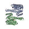
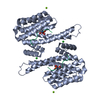
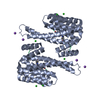
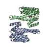
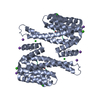
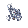
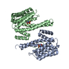
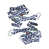
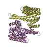
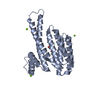
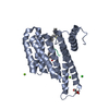
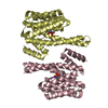
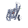
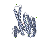
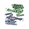
- PDB-6bcr: Complex of 14-3-3 theta with an IRSp53 peptide phosphorylated at T340 -
+ Open data

- Basic information
| Entry | Database: PDB / ID: 6bcr | ||||||
|---|---|---|---|---|---|---|---|
| Title | Complex of 14-3-3 theta with an IRSp53 peptide phosphorylated at T340 | ||||||
 Components Components |
| ||||||
 Keywords Keywords | SIGNALING PROTEIN / phosphate binding protein / protein complex / cytoskeleton regulation / cell motility | ||||||
| Function / homology |  Function and homology information Function and homology informationneuron projection branch point / plasma membrane organization / actin crosslink formation / cadherin binding involved in cell-cell adhesion / negative regulation of monoatomic ion transmembrane transport / regulation of modification of postsynaptic actin cytoskeleton / cytoskeletal adaptor activity / positive regulation of dendritic spine morphogenesis / protein localization to synapse / proline-rich region binding ...neuron projection branch point / plasma membrane organization / actin crosslink formation / cadherin binding involved in cell-cell adhesion / negative regulation of monoatomic ion transmembrane transport / regulation of modification of postsynaptic actin cytoskeleton / cytoskeletal adaptor activity / positive regulation of dendritic spine morphogenesis / protein localization to synapse / proline-rich region binding / cellular response to L-glutamate / dendritic spine cytoplasm / small GTPase-mediated signal transduction / positive regulation of actin filament polymerization / neuron projection terminus / dendrite development / CDC42 GTPase cycle / Regulation of localization of FOXO transcription factors / actin filament bundle assembly / RAC3 GTPase cycle / Activation of BAD and translocation to mitochondria / RHO GTPases Activate WASPs and WAVEs / protein targeting / SARS-CoV-2 targets host intracellular signalling and regulatory pathways / postsynaptic cytosol / postsynaptic density, intracellular component / Chk1/Chk2(Cds1) mediated inactivation of Cyclin B:Cdk1 complex / SARS-CoV-1 targets host intracellular signalling and regulatory pathways / RHO GTPases activate PKNs / presynaptic cytosol / ruffle / axonogenesis / RAC1 GTPase cycle / 14-3-3 protein binding / substantia nigra development / excitatory synapse / secretory granule / cellular response to epidermal growth factor stimulus / positive regulation of excitatory postsynaptic potential / transcription coregulator binding / dendritic shaft / regulation of actin cytoskeleton organization / TP53 Regulates Metabolic Genes / synaptic membrane / FCGR3A-mediated phagocytosis / filopodium / Translocation of SLC2A4 (GLUT4) to the plasma membrane / adherens junction / PDZ domain binding / Regulation of actin dynamics for phagocytic cup formation / regulation of synaptic plasticity / VEGFA-VEGFR2 Pathway / Schaffer collateral - CA1 synapse / insulin receptor signaling pathway / intracellular protein localization / regulation of cell shape / lamellipodium / scaffold protein binding / microtubule / transmembrane transporter binding / protein domain specific binding / focal adhesion / negative regulation of DNA-templated transcription / neuronal cell body / synapse / glutamatergic synapse / signal transduction / protein-containing complex / extracellular exosome / nucleoplasm / membrane / identical protein binding / nucleus / plasma membrane / cytosol / cytoplasm Similarity search - Function | ||||||
| Biological species |  Homo sapiens (human) Homo sapiens (human) | ||||||
| Method |  X-RAY DIFFRACTION / SYNCHROTRON / MOLECULAR REPLACEMENT / molecular replacement / Resolution: 1.986 Å X-RAY DIFFRACTION / SYNCHROTRON / MOLECULAR REPLACEMENT / molecular replacement / Resolution: 1.986 Å | ||||||
 Authors Authors | Kast, D.J. / Dominguez, R. | ||||||
| Funding support |  United States, 1items United States, 1items
| ||||||
 Citation Citation | Journal: Nat Commun / Year: 2019 Title: Mechanism of IRSp53 inhibition by 14-3-3. Authors: Kast, D.J. / Dominguez, R. | ||||||
| History |
|
- Structure visualization
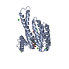
| Structure viewer | Molecule: MolmilJmol/JSmol |
|---|
- Downloads & links
-Download
| PDBx/mmCIF format | 6bcr.cif.gz | 390.5 KB | Display | PDBx/mmCIF format |
|---|---|---|---|---|
| PDB format | pdb6bcr.ent.gz | 322.5 KB | Display | PDB format |
| PDBx/mmJSON format | 6bcr.json.gz | Tree view | PDBx/mmJSON format | |
| Others |  Other downloads Other downloads |
-Validation report
| Arichive directory | https://data.pdbj.org/pub/pdb/validation_reports/bc/6bcrftp://data.pdbj.org/pub/pdb/validation_reports/bc/6bcr | HTTPS FTP |
|---|
-Related structure data
| Related structure data | 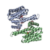 6bcyC 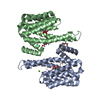 6bd1C 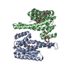 6bd2C 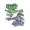 6bqtC  2br9S S: Starting model for refinement C: citing same article ( |
|---|---|
| Similar structure data |

-Links
 PDBj
PDBj


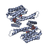
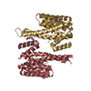
- Assembly
| Deposited unit | 
| ||||||||
|---|---|---|---|---|---|---|---|---|---|
| 1 | 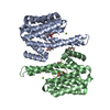
| ||||||||
| 2 | 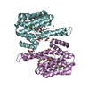
| ||||||||
| Unit cell |
|
-Components
-Protein / Protein/peptide , 2 types, 8 molecules ABEFCDGH
| #1: Protein | Mass: 27795.234 Da / Num. of mol.: 4 Source method: isolated from a genetically manipulated source Source: (gene. exp.) Homo sapiens (human) / Gene: YWHAQ / Plasmid: pTYB11 / Production host:  #2: Protein/peptide | Mass: 1648.709 Da / Num. of mol.: 4 / Source method: obtained synthetically / Source: (synth.) Homo sapiens (human) / References: UniProt: Q9UQB8*PLUS |
|---|
-Non-polymers , 6 types, 533 molecules 


| #3: Chemical | ChemComp-MG /  Mass: 24.305 Da / Num. of mol.: 5 / Source method: obtained synthetically / Formula: Mg Mass: 24.305 Da / Num. of mol.: 5 / Source method: obtained synthetically / Formula: Mg#4: Chemical | ChemComp-PEG /  Mass: 106.120 Da / Num. of mol.: 5 / Source method: obtained synthetically / Formula: C4H10O3 Mass: 106.120 Da / Num. of mol.: 5 / Source method: obtained synthetically / Formula: C4H10O3#5: Chemical |  Mass: 238.278 Da / Num. of mol.: 2 / Source method: obtained synthetically / Formula: C10H22O6 / Comment: precipitant*YM Mass: 238.278 Da / Num. of mol.: 2 / Source method: obtained synthetically / Formula: C10H22O6 / Comment: precipitant*YM#6: Chemical |  Mass: 62.068 Da / Num. of mol.: 2 / Source method: obtained synthetically / Formula: C2H6O2 Mass: 62.068 Da / Num. of mol.: 2 / Source method: obtained synthetically / Formula: C2H6O2#7: Chemical |  Mass: 100.040 Da / Num. of mol.: 2 / Source method: obtained synthetically / Formula: C2H3F3O Mass: 100.040 Da / Num. of mol.: 2 / Source method: obtained synthetically / Formula: C2H3F3O#8: Water | ChemComp-HOH / | Mass: 18.015 Da / Num. of mol.: 517 / Source method: isolated from a natural source / Formula: H2O |
|---|
-Details
| Has protein modification | Y |
|---|
-Experimental details
-Experiment
| Experiment | Method: X-RAY DIFFRACTION / Number of used crystals: 1 |
|---|
- Sample preparation
| Crystal | Density Matthews: 2.53 Å3/Da / Density % sol: 51.3 % |
|---|---|
| Crystal grow | Temperature: 291.15 K / Method: vapor diffusion, hanging drop / pH: 7 / Details: 0.15 M Magnesium Formate, 18% PEG3350, 4% TFE |
-Data collection
| Diffraction | Mean temperature: 100 K |
|---|---|
| Diffraction source | Source: SYNCHROTRON / Site: NSLS / Beamline: X6A / Wavelength: 1 Å |
| Detector | Type: ADSC QUANTUM 270 / Detector: CCD / Date: Jun 17, 2012 / Details: Oxford Danfysik toroidal focusing mirror |
| Radiation | Monochromator: channel cut monochromator / Protocol: SINGLE WAVELENGTH / Monochromatic (M) / Laue (L): M / Scattering type: x-ray |
| Radiation wavelength | Wavelength: 1 Å / Relative weight: 1 |
| Reflection | Resolution: 1.986→31.29 Å / Num. obs: 76071 / % possible obs: 95.7 % / Redundancy: 3.4 % / Biso Wilson estimate: 32.72 Å2 / CC1/2: 0.999 / Rmerge(I) obs: 0.042 / Net I/σ(I): 21.8 |
| Reflection shell | Resolution: 1.986→2.06 Å / Redundancy: 2.7 % / Rmerge(I) obs: 0.379 / Mean I/σ(I) obs: 3.5 / Num. unique obs: 6527 / CC1/2: 0.888 / % possible all: 92 |
-Phasing
| Phasing | Method: molecular replacement |
|---|
- Processing
| Software |
| ||||||||||||||||||||||||||||||||||||||||||||||||||||||||||||||||||||||||||||||||||||||||||||||||||||||||||||||||||||||||||||||||||||||||||||||||||||||||||||||||||||||||||||||||||||||||||||||||||||
|---|---|---|---|---|---|---|---|---|---|---|---|---|---|---|---|---|---|---|---|---|---|---|---|---|---|---|---|---|---|---|---|---|---|---|---|---|---|---|---|---|---|---|---|---|---|---|---|---|---|---|---|---|---|---|---|---|---|---|---|---|---|---|---|---|---|---|---|---|---|---|---|---|---|---|---|---|---|---|---|---|---|---|---|---|---|---|---|---|---|---|---|---|---|---|---|---|---|---|---|---|---|---|---|---|---|---|---|---|---|---|---|---|---|---|---|---|---|---|---|---|---|---|---|---|---|---|---|---|---|---|---|---|---|---|---|---|---|---|---|---|---|---|---|---|---|---|---|---|---|---|---|---|---|---|---|---|---|---|---|---|---|---|---|---|---|---|---|---|---|---|---|---|---|---|---|---|---|---|---|---|---|---|---|---|---|---|---|---|---|---|---|---|---|---|---|---|---|
| Refinement | Method to determine structure: MOLECULAR REPLACEMENT Starting model: 2BR9 Resolution: 1.986→31.288 Å / SU ML: 0.22 / Cross valid method: FREE R-VALUE / σ(F): 1.96 / Phase error: 25.81
| ||||||||||||||||||||||||||||||||||||||||||||||||||||||||||||||||||||||||||||||||||||||||||||||||||||||||||||||||||||||||||||||||||||||||||||||||||||||||||||||||||||||||||||||||||||||||||||||||||||
| Solvent computation | Shrinkage radii: 0.9 Å / VDW probe radii: 1.11 Å | ||||||||||||||||||||||||||||||||||||||||||||||||||||||||||||||||||||||||||||||||||||||||||||||||||||||||||||||||||||||||||||||||||||||||||||||||||||||||||||||||||||||||||||||||||||||||||||||||||||
| Displacement parameters | Biso max: 135.83 Å2 / Biso mean: 46.5178 Å2 / Biso min: 17.43 Å2 | ||||||||||||||||||||||||||||||||||||||||||||||||||||||||||||||||||||||||||||||||||||||||||||||||||||||||||||||||||||||||||||||||||||||||||||||||||||||||||||||||||||||||||||||||||||||||||||||||||||
| Refinement step | Cycle: final / Resolution: 1.986→31.288 Å
| ||||||||||||||||||||||||||||||||||||||||||||||||||||||||||||||||||||||||||||||||||||||||||||||||||||||||||||||||||||||||||||||||||||||||||||||||||||||||||||||||||||||||||||||||||||||||||||||||||||
| Refine LS restraints |
| ||||||||||||||||||||||||||||||||||||||||||||||||||||||||||||||||||||||||||||||||||||||||||||||||||||||||||||||||||||||||||||||||||||||||||||||||||||||||||||||||||||||||||||||||||||||||||||||||||||
| LS refinement shell | Refine-ID: X-RAY DIFFRACTION / Rfactor Rfree error: 0 / Total num. of bins used: 27
|
